
Image
|
Figure Caption
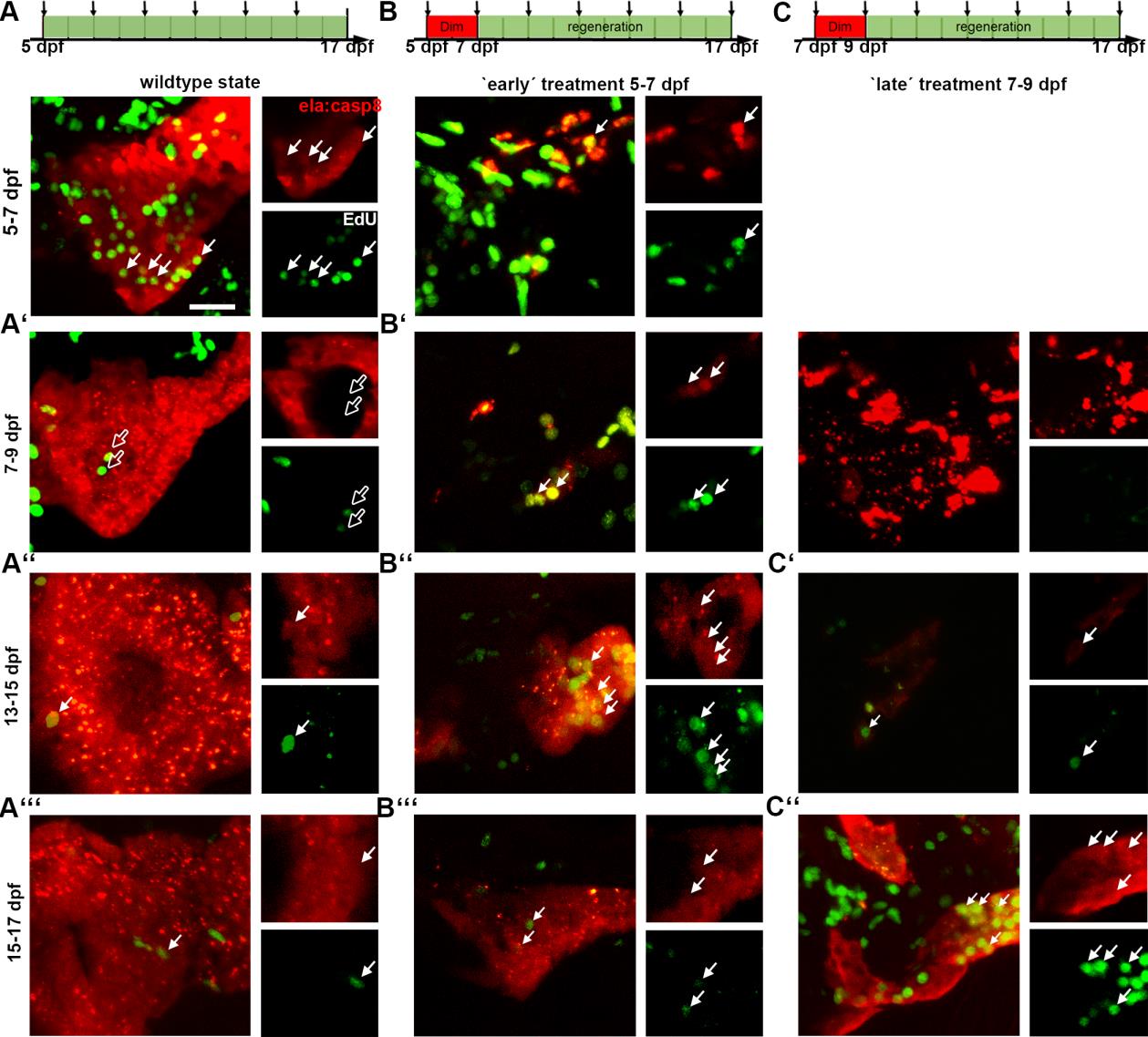
Fig. S3
Dynamics of exocrine development and regeneration. Confocal projections and single plane images of the untreated and regenerating anterior portion of the exocrine pancreas of Tg(ela:casp8) stained for EdU. Larvae were treated with EdU for 48 hours before harvest. A: the exocrine pancreas in wildtype-state, E2Crimson positive exocrine cells and EdU incorporation at 7, 9, 15 and 17 dpf B: larvae treated with 5μM Dim from 5 to 7 dpf and injected with EdU at different time points. C: larvae treated with 5μM Dim from 7 to 9 dpf and injected with EdU. Scale bar: 20μm.
Acknowledgments
This image is the copyrighted work of the attributed author or publisher, and
ZFIN has permission only to display this image to its users.
Additional permissions should be obtained from the applicable author or publisher of the image.
Full text @ Dis. Model. Mech.