
Image
|
Figure Caption
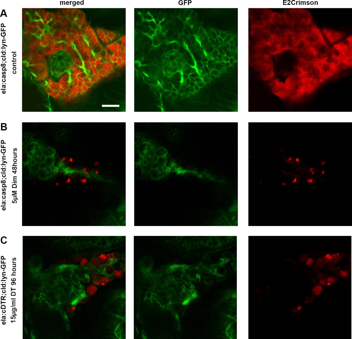
Fig. S1
Loss of cell membranes of targeted exocrine cells. Tg(cld:lyn-GFP) was used to label cell membranes. Confocal projections of A, B: Tg(ela:casp8;ela:E2crimson) control and ablated larvae at 7 dpf. C: Tg(ela:DTR;ela:E2crimson) ablated larvae at 9 dpf. In B and C, E2crimson signal is mostly located in debris not associated with cell membranes and thus does not label intact exocrine cells after treatment. Scale bar: 20μm.
Acknowledgments
This image is the copyrighted work of the attributed author or publisher, and
ZFIN has permission only to display this image to its users.
Additional permissions should be obtained from the applicable author or publisher of the image.
Full text @ Dis. Model. Mech.