
|
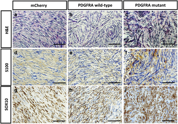
Fig. 3
PDGFRA-induced zebrafish MPNST tumors exhibit similar histology to that of human MPNSTs. (a–c) The histopathology after H&E staining of the zebrafish MPNSTs was identical in the nf1a+/−; nf1b−/−; p53m/m zebrafish when either PDGFRA wild-type or mutant proteins were overexpressed. In each genotype, the zebrafish MPNST histopathology was very similar to that of human MPNSTs. The zebrafish MPNSTs comprise spindle cells that stack into short fascicles, typically with a whirling organization pattern. MPNST cells have long serpentine-like nuclei and are spindle-shaped. (d–i) Markers of cells of neural crest origin were expressed by the zebrafish MPNST tumors, such as S100 (d–f) and Sox10 (g–i). S100 was detected in the cell membrane and nucleus, whereas Sox10 was detected in the nucleus. Scale bar=200 μm.