
|
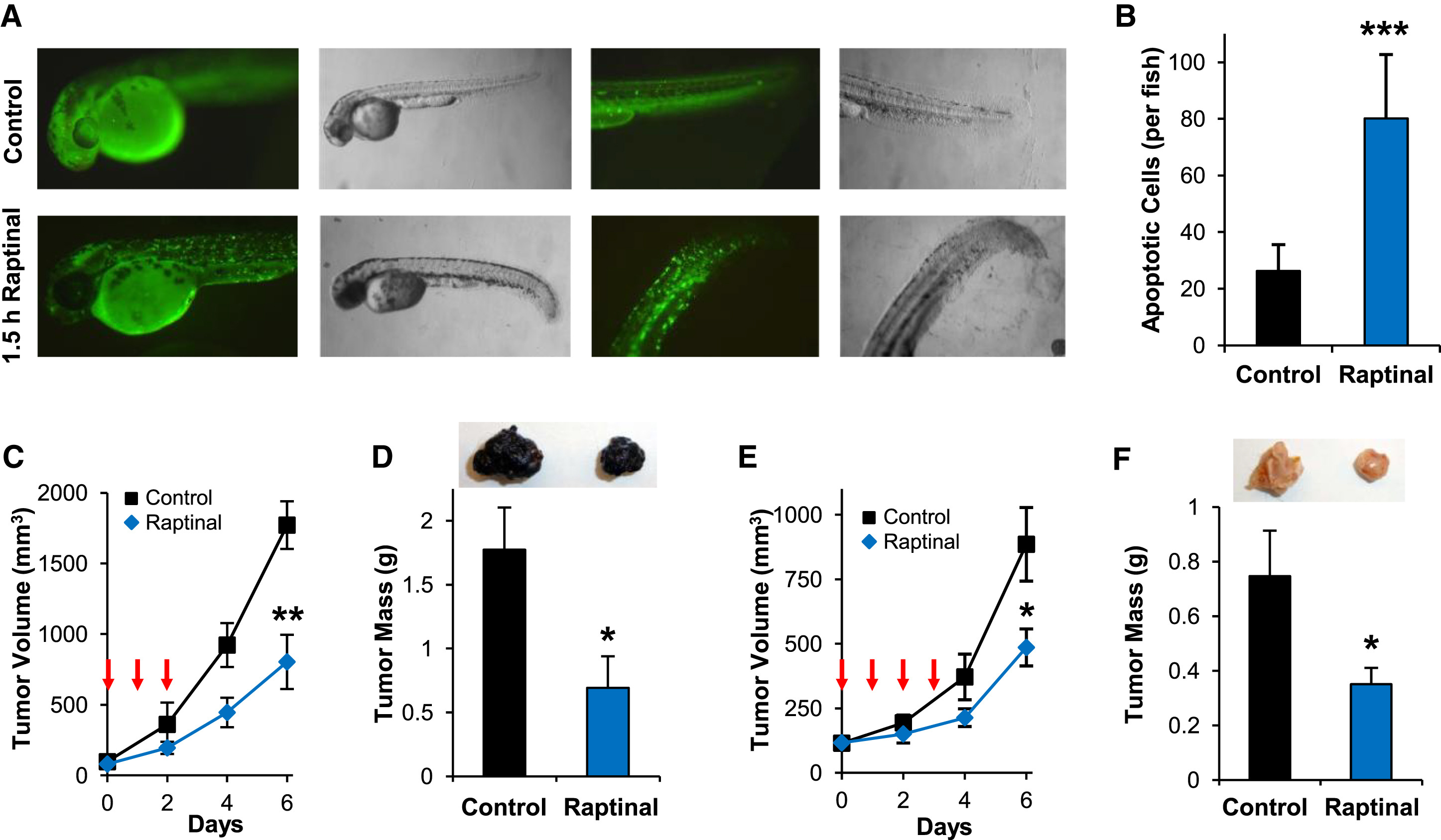
Fig. 3
Raptinal Exhibits Activity In Vivo
(A) Zebrafish embryos expressing secreted annexin V-YFP exhibit pronounced punctate YFP signal indicating phosphatidylserine externalization following 1.5 hr of treatment with 10 μM Raptinal.
(B) Quantification of apoptotic cells in Raptinal- versus DMSO-treated zebrafish under the conditions in (A). Data represent the mean ± SD (n = 5 and n = 7 embryos for Raptinal and DMSO, respectively; ∗∗∗p value <0.001).
(C and D) Raptinal inhibits subcutaneous B16-F10 melanoma tumor growth in vivo as measured by tumor volume (∗∗p value < 0.005) in (C) and tumor mass after tumor excision in (D) (∗p value <0.05).
(E and F) Raptinal inhibits subcutaneous 4T1 breast cancer tumor growth in vivo as measured by tumor volume (∗p value <0.05) in (E) and tumor mass after tumor excision (∗p value <0.05) in (F).
Arrows in (C) and (E) indicate intraperitoneal Raptinal administration at 20 mg/kg once a day. Tumor images in (D) and (F) are representative of tumor size at the conclusion of the studies. Data in (C)–(F) represent the mean ± SEM (n = 7 mice/group). See also Figure S3.