
Fig. 1
- ID
- ZDB-IMAGE-170413-53
- Genes
- Publication
- Carroll et al., 2014 - Estrogen defines the dorsal-ventral limit of VEGF regulation to specify the location of the hemogenic endothelial niche
- All Figures
- Figures for Carroll et al., 2014
|
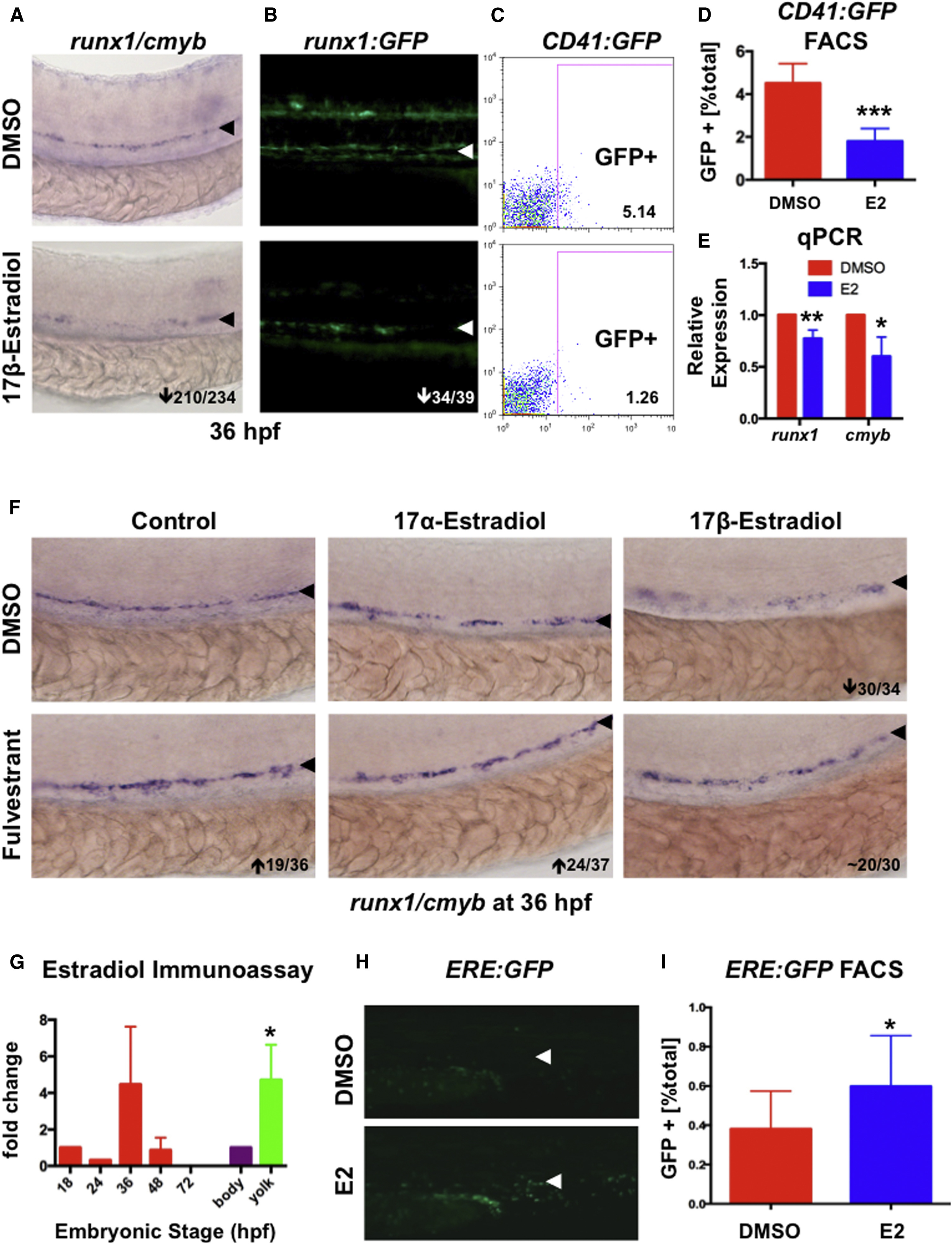
Fig. 1
Exposure to E2 Impairs Formation of HSPCs in the AGM
(A) E2 exposure from five somites to 36 hpf decreased runx1/cmyb expression in the AGM (210/234).
(B) E2 diminished runx1:GFP+ cells in the AGM (34/39).
(C) FACS analysis of CD41:GFP embyros (representative samples shown) confirmed that E2 reduced HSCs.
(D) CD41:GFP was significantly decreased by E2 (n = 20, two-tailed t test, p < 0.001; error bars indicate SD).
(E) qPCR quantified reduced runx1 and cmyb expression by E2 (mean of triplicate experiments ± SEM; one-tailed t test runx∗∗p < 0.01, cmyb∗p < 0.05).
(F) The pan-Esr antagonist FULV blocked the effect of E2; the isomer 17α-estradiol (10 μM) had no effect (n ≥ 30/treatment).
(G) Estradiol EIA revealed that endogenous estrogens are present during hematopoiesis; E2 was enriched 4.7-fold in the yolk versus body at 18 hpf (mean of triplicate experiments ± SEM; two-tailed t test, ∗p < 0.05).
(H) ERE:GFP fish show estrogenic activity in the AGM.
(I) GFP expression (as seen in H) was increased 1.57-fold upon E2 exposure (n = 15, two-tailed t test, p < 0.05; error bars indicate SD).
The number of embryos with altered expression over the total number analyzed is shown (bottom right).
Reprinted from Developmental Cell, 29, Carroll, K.J., Esain, V., Garnaas, M.K., Cortes, M., Dovey, M.C., Nissim, S., Frechette, G.M., Liu, S.Y., Kwan, W., Cutting, C.C., Harris, J.M., Gorelick, D.A., Halpern, M.E., Lawson, N.D., Goessling, W., North, T.E., Estrogen defines the dorsal-ventral limit of VEGF regulation to specify the location of the hemogenic endothelial niche, 437-53, Copyright (2014) with permission from Elsevier. Full text @ Dev. Cell