
|
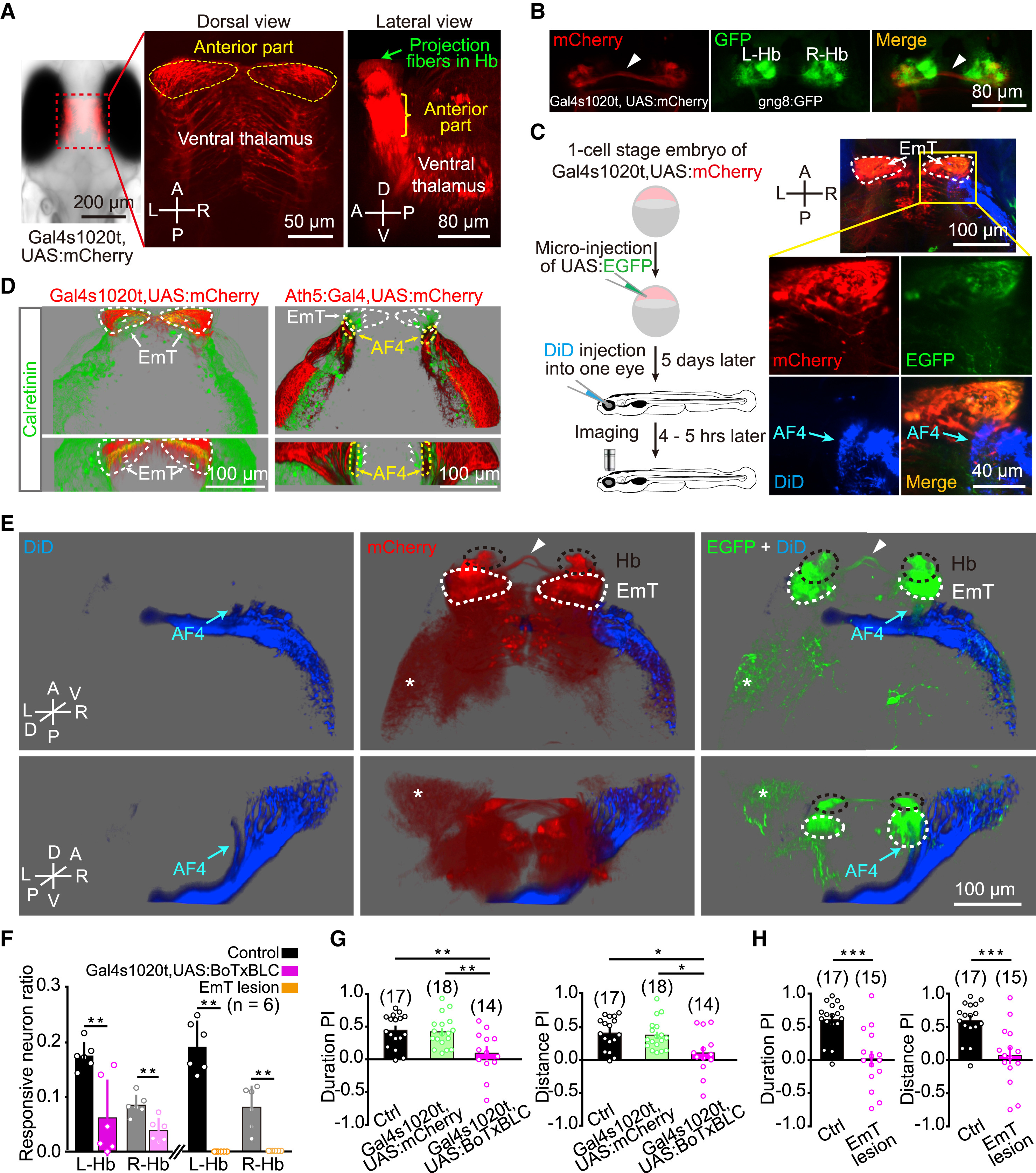
Fig. 5
EmT Relay Visual Inputs from AF4-Projecting RGCs to L-dHb and Are Critical for Light-Preference Behavior
(A) Projected confocal images of a 6-dpf Tg(Gal4s1020t,UAS:mCherry) larva showing mCherry expression in the ventral thalamus, in particular its anterior part (i.e., the eminentia thalami, EmT). The green arrow in the right panel points to mCherry-positive fibers in the Hb region.
(B) Projected confocal images of a 6-dpf Tg(Gal4s1020t,UAS:mCherry,gng8:GFP) larva showing mCherry signal overlaps GFP signal in the dHb. Arrowhead, Hb commissure.
(C) Sparse labeling of EmT neurons by transient GFP expression and dense labeling of RGC axons by DiD injection into one eye in Tg(Gal4s1020t,UAS:mCherry) larvae. Left: experimental diagram; right: example of projected confocal images showing neurons in the EmT region are contacted by DiD-positive RGC axons (blue) in the arborization field 4 (AF4, arrow), a retinorecipient brain region.
(D) Projected confocal images (top: dorsal view; bottom: anterior view) of immunostaining of calretinin, a marker of EmT neurons, in Tg(Gal4s1020t,UAS:mCherry) (left) and Tg(Ath5:Gal4,UAS:mCherry) (right) larvae. White arrowheads, calretinin-positive cells in the EmT area.
(E) Three-dimensional representation of the case shown in (C). DiD-labeled RGC axons are in blue, EGFP-expressing EmT neurons are in green, and mCherry-expressing thalamic neurons are in red. EmT neurons are contacted by RGC axons at the AF4 and send projection fibers to the Hb. White arrowhead, Hb commissure; asterisk, thalamic fibers in the optic tectum.
(F) Ratio of visually responsive neurons in both the L-Hb and R-Hb of control, Tg(Gal4s1020t,UAS:BoTxBLC-GFP), and EmT lesion larvae at 6 dpf. Data from each larva were obtained from the whole five layers of the Hb.
(G) Effects of synaptic blockade of the ventral thalamus (including the EmT) on light-preference behavior. WT control (in black), Tg(Gal4s1020t,UAS:mCherry) (in green), and Tg(Gal4s1020t,UAS:BoTxBLC-GFP) (in pink) larvae at 6 dpf were used.
(H) Effects of bilateral EmT lesion on light-preference behavior. Two-photon laser-induced lesion was performed on 6-dpf Tg(Gal4s1020t,UAS:GFP) larvae.
The numbers in the parentheses represent the numbers of larvae examined (F, G, and H). Error bars, SEM; ∗p < 0.05, ∗∗p < 0.01, ∗∗∗p < 0.001 (unpaired two-tailed Student’s t test for comparison between control and Tg(Gal4s1020t,UAS:BoTxBLC-GFP) groups in F; Mann-Whitney test for comparison between control and EmT lesion groups in F; one-way ANOVA for G; unpaired two-tailed Student’s t test for H).
See also Figures S4 and S5.