
|
Fig. 1
A Balbiani Body Is a Non-Membrane-Bound Compartment Packed with Membranous Organelles
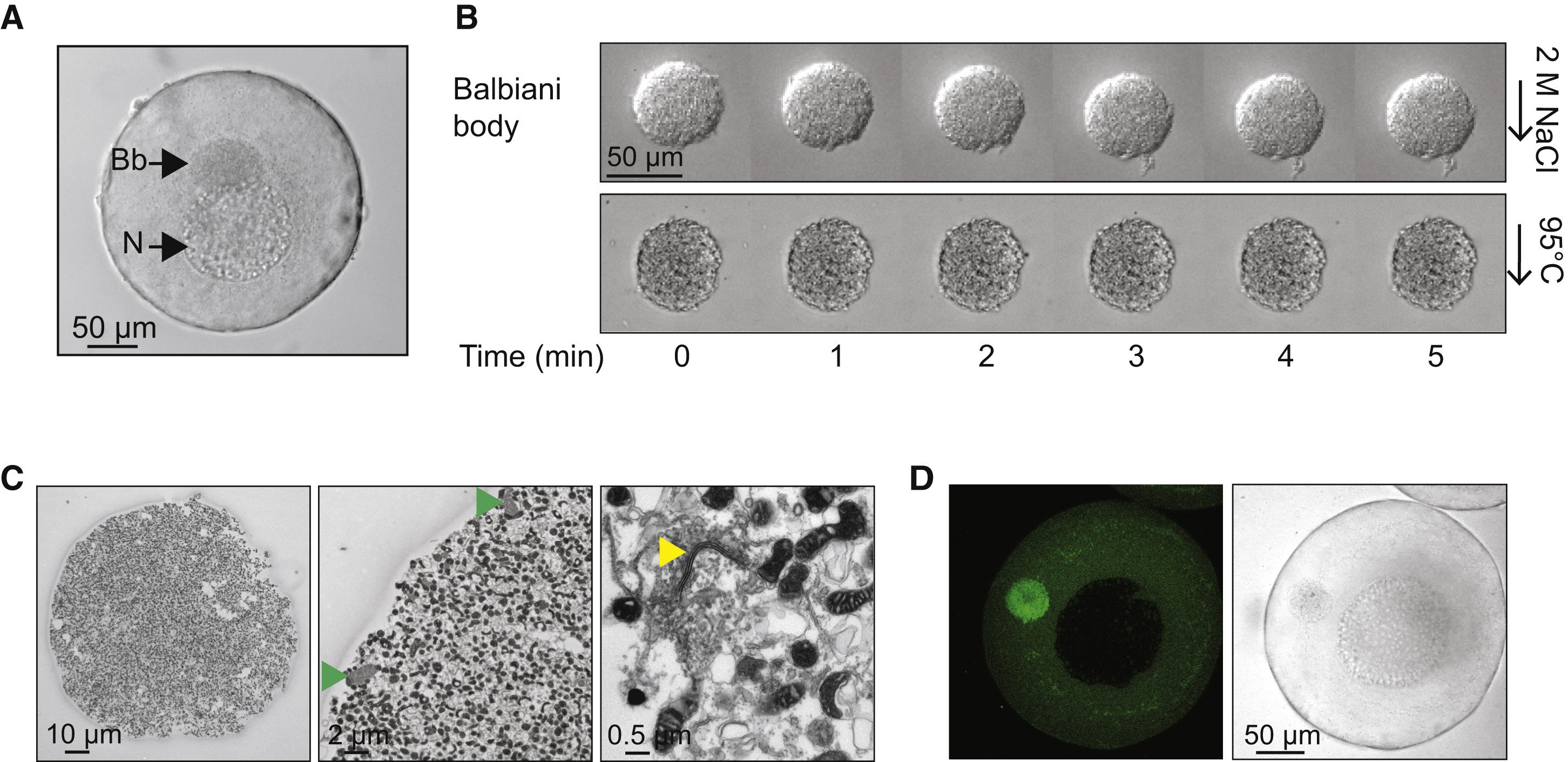
(A) Phase contrast image of a stage I Xenopus laevis oocyte. Bb, Balbiani body; N, nucleus or germinal vesicle.
(B) Balbiani body immobilized in perfusion chambers. 2 M NaCl (first panel) or 95°C 50 mM HEPES, 100 mM KCl (pH 7.6) buffer (second panel) was perfused into the chambers.
(C) Thin-section electron microscope (EM) images of isolated Balbiani bodies from stage I Xenopus oocytes. Mitochondria (dark spots), RNP particles (green arrow head), and Golgi stacks (yellow arrow head) are clearly visible.
(D) Stage I oocytes were incubated in 10 μM Thioflavin T in 1× MMR for 10 min and washed twice with 1× MMR.
See also Figure S1, Table S1, and Movie S1.
Reprinted from Cell, 166, Boke, E., Ruer, M., Wühr, M., Coughlin, M., Lemaitre, R., Gygi, S.P., Alberti, S., Drechsel, D., Hyman, A.A., Mitchison, T.J., Amyloid-like Self-Assembly of a Cellular Compartment, 637-50, Copyright (2016) with permission from Elsevier. Full text @ Cell