
Fig. 5
- ID
- ZDB-IMAGE-170314-5
- Genes
- Publication
- Williams et al., 2017 - Cyp1b1 Regulates Ocular Fissure Closure Through a Retinoic Acid-Independent Pathway
- All Figures
- Figures for Williams et al., 2017
|
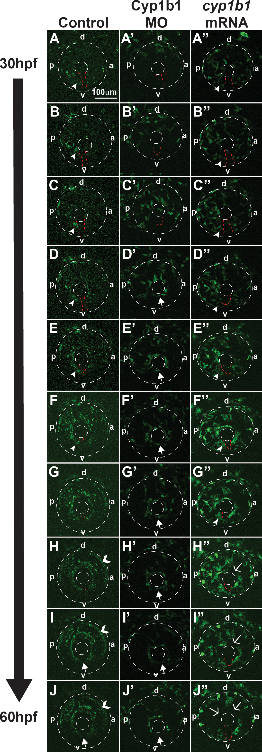
Fig. 5
Cyp1b1 regulates ocular neural crest migration. Time-lapse imaging of Tg(foxd3:EGFP) control embryos from 30 to 60 hpf (A–J) showed that foxd3-positive cells entered into the anterior chamber between the surface ectoderm and optic cup, with more cells localized to the dorsal (d)–posterior (p) quadrant (E–H) compared with the anterior (a) and ventral (v) areas. In addition, foxd3-positive cells migrated adjacent to and through the ocular fissure (A–F, white arrowhead denotes migrating neural crest, red dashed line demarcates ocular fissure). At 60 hpf, foxd3-positive cells completely encircled the lens (I, J, closed arrows), indicating the closure of the fissure. In addition, at 60 hpf, foxd3 was also expressed in photoreceptors (H–J, open arrowhead). In Cyp1b1 MO knockdown embryos, Foxd3-positive cells travelled between the surface ectoderm and optic cup in the dorsal quadrants (A'–E'), but there were few cells adjacent to and in the ocular fissure. Foxd3-positive cells encircled the lens prior to controls (D'–H' versus D–H), indicating the premature closure of the fissure. At 60 hpf, there were fewer foxd3-positive cells in the eye (J'). In embryos overexpressing cyp1b1, ocular neural crest migration was less organized, as foxd3-positive cells were located throughout the posterior half of the eye (A''–G''). Further, the ocular fissure remained open and prevented the continuous coalescence of neural crest cells around the lens (H''–J'', open arrows), resulting in fewer neural crest cells at 60 hpf (J'').