
|
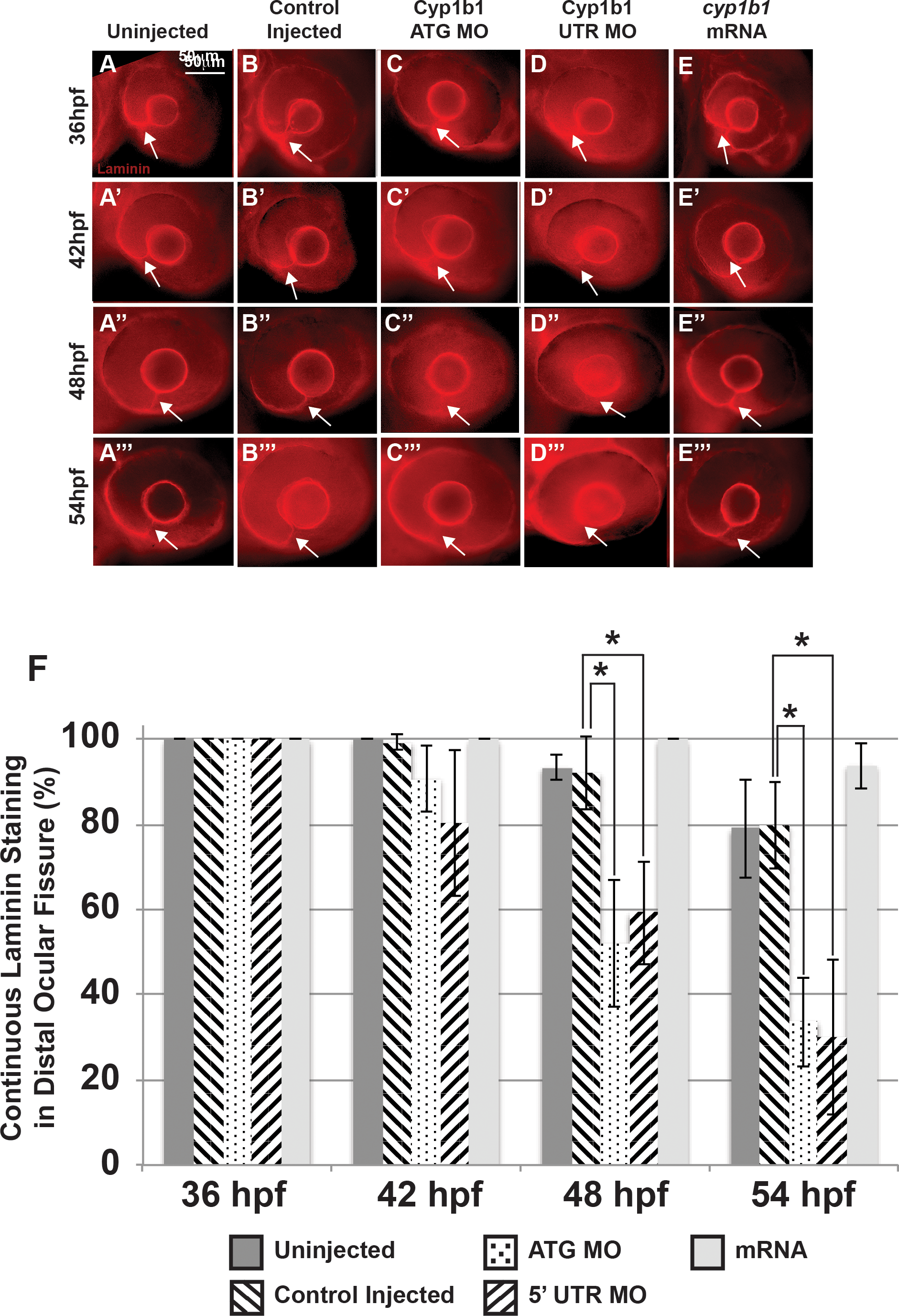
Fig. 4
Alterations in the basement membrane integrity as a result of decreased or increased Cyp1b1. Whole-mount laminin staining showed that in uninjected (A–A''') and control-injected (B–B''') embryos, a continuous basement membrane (arrows) was evident in the distal ocular fissure at 36, 42, 48, and 54 hpf. Cyp1b1 ATG (C–C''') and 5′ UTR (D–D''') MO knockdown showed discontinuous staining of laminin at 48 (C'', D'') and 54 hpf (C''', D'''), suggesting premature basement membrane breakdown. In cyp1b1 mRNA-injected embryos, laminin staining remained prominent within the fissure at all time points (E–E'''). The percentage of embryos with continuous laminin staining in the distal ocular fissure at 36, 42, 48, and 54 hpf (G), (*P < 0.001).