
|
Fig. 1
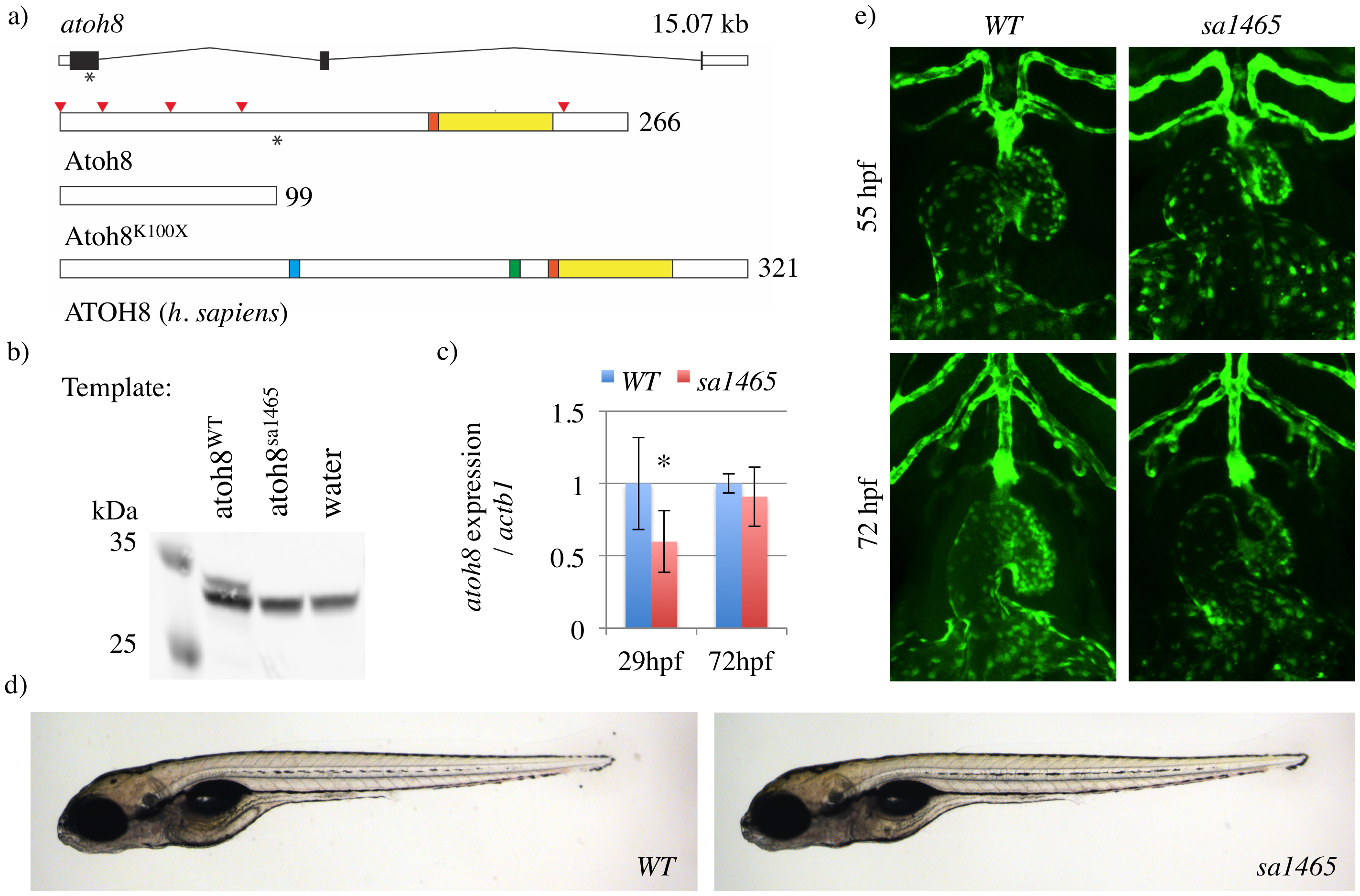
Atoh8sa1465/sa1465 mutants are morphologically normal, with correct heart looping.
a) Structure of the zebrafish atoh8 locus and Atoh8 protein. The positions of the A > T substitution in the atoh8sa1465 allele, and of Lysine 100 in Atoh8 protein, are indicated with asterisks. The red arrowheads mark the positions of each Methionine in the Atoh8 protein, and the basic (orange) and HLH (yellow) domains are indicated. The truncated protein Atoh8K100X is the predicted product of atoh8sa1465. Human ATOH8 contains additional proline-rich (blue) and serine-rich (green) domains. b) Atoh8 Western blot on products from in vitro transcription/translation reactions (TNT Quick) using atoh8WT/WT and atoh8sa1465/sa1465 as templates. Predicted size of Atoh8 = 29.8 kDa. Note that there is a strong nonspecific band at ~31 kDa. The full length membrane can be viewed in S1 Fig. c) qPCR on atoh8WT/WT and atoh8sa1465/sa1465 embryos (hereafter labelled as 'WT' and 'sa1465', respectively). * p = 0.028. d) WT and sa1465 embryos at 5 days postfertilisation, showing normal overall body morphology and swimbladder inflation. e) Confocal z-stacks showing WT and sa1465 embryo heart morphology.