
Image
|
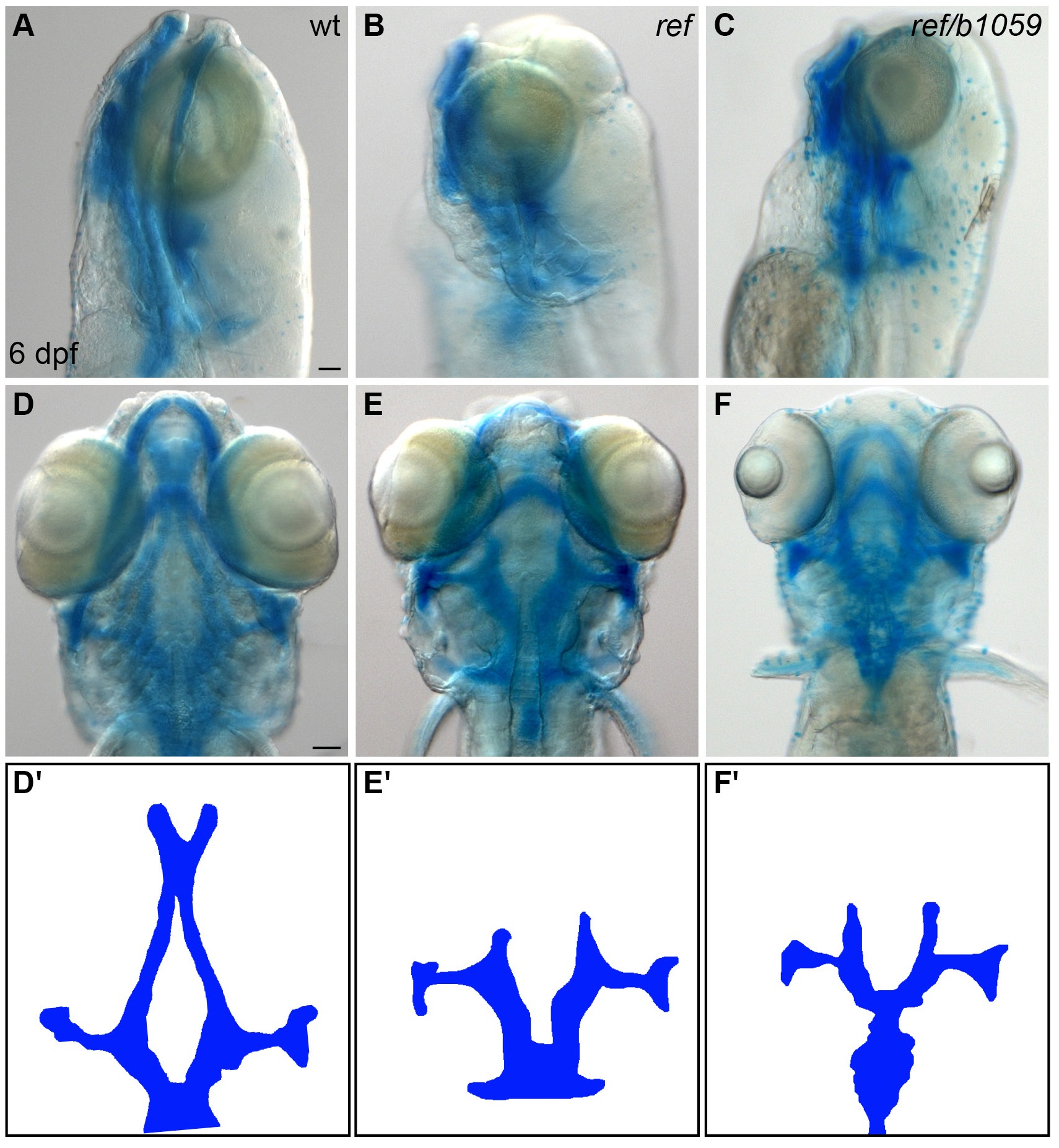
Figure Caption
Fig. 2 S1
ref mutants display craniofacial defects.
(A–F) Lateral (A–C) and ventral (D–F) views of embryos at 6 days post-fertilization (dpf), stained with Alcian blue to show the dorsal and ventral cartilage in the head. Embryos homozygous for ref (B,E; n = 9/9) or transheterozgous for ref and b1059 (C,F; n = 5/6) display similar dorsal cartilage formation defects. Specifically, the dorsal ethmoid plate is diminished or absent in these mutants. Scale bars: 60 μm. (D'–F') Cartoons illustrate the dorsal cartilage structures shown in D–F.
Figure Data
Acknowledgments
This image is the copyrighted work of the attributed author or publisher, and
ZFIN has permission only to display this image to its users.
Additional permissions should be obtained from the applicable author or publisher of the image.
Full text @ Elife