
|
Fig. 1
Synchronous Spontaneous Activity of Dopaminergic Cell Subgroups in the Diencephalon
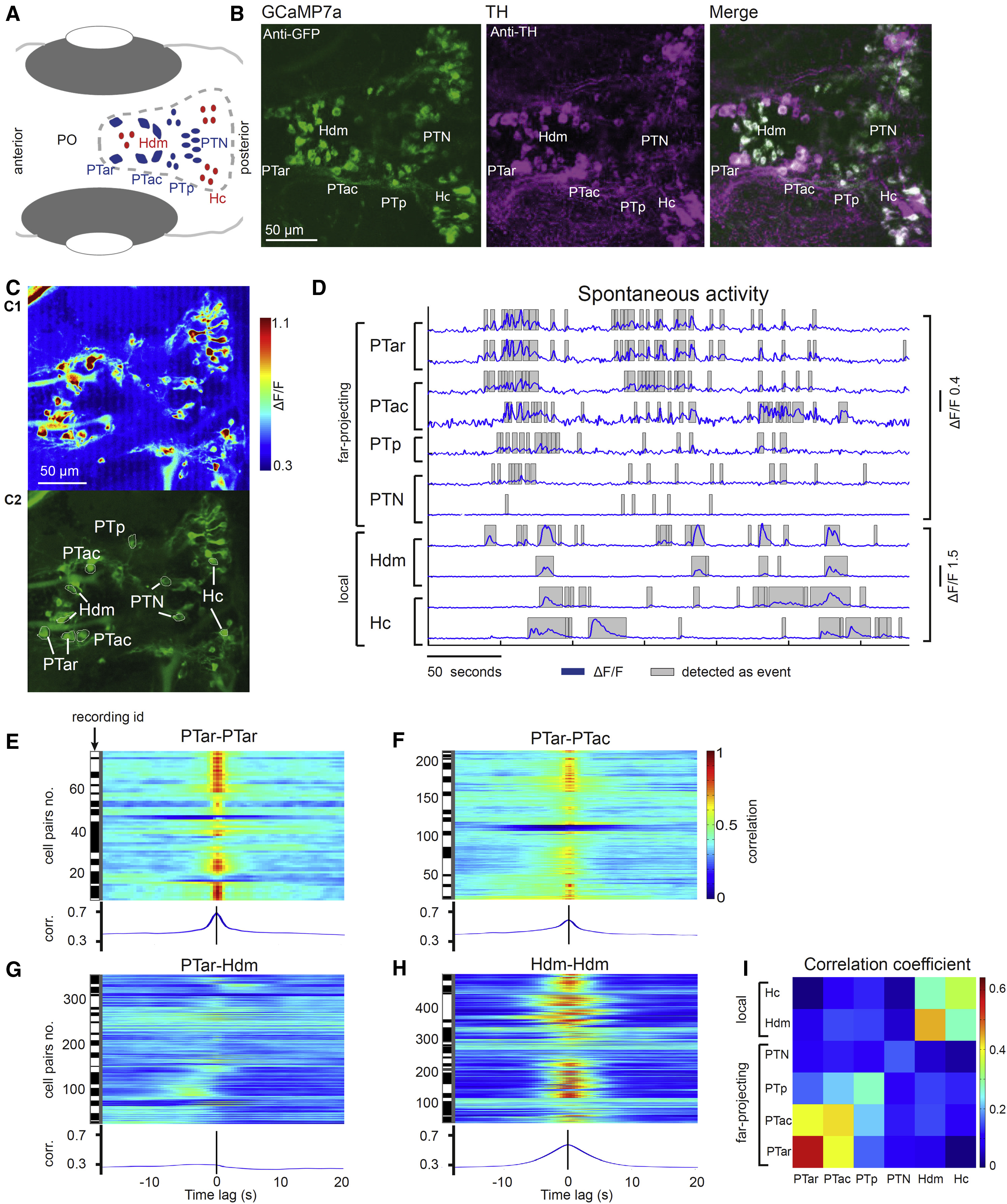
(A) Schematic of diencephalic dopaminergic subgroups. Far-projecting posterior tubercular neurons are marked in blue, and predominantly locally projecting hypothalamic neurons are marked in red. PO, preoptic area.
(B) Colocalization of tyrosine hydroxylase (TH) and GCaMP7a. Anti-GFP (detects GCaMP7a) and anti-TH immunofluorescence stain of a Tg(th:Gal4VP16)m1233, Tg(UAS:GCaMP7a)zf415Tg larva (ventral diencephalon, 4 dpf, z-projections).
(C) In vivo calcium imaging of dopaminergic neurons in the diencephalon. (C1) Mean ΔF/F heatmap of a calcium image time series. (C2) Standard deviation time series projection visualizing active, expressing neurons was solely used to identify neurons for further analysis.
(D) Calcium signals (ΔF/F) of the marked cells of different dopaminergic subgroups shown in (C2).
(E–H) Cross-correlations of spontaneous calcium activity in cell pairs of the same dopaminergic subgroup (E and H) and different subgroups (F and G). The black and white bars each indicate different recordings, i.e., all cell pairs from one contiguous black or white bar belong to the same recording. Rostral far-projecting neurons in PTar and PTac show synchronous activity (E and F), whereas PTar activity is only weakly correlated with activity in local Hdm dopaminergic neurons (G). Within Hdm, neural activity is synchronous (H). corr., correlation.
(I) Mean correlation coefficients of all cell pairs within and across dopaminergic subgroups. The activity is only weakly correlated (blue) between far-projecting (PTar and PTac) and local hypothalamic groups (Hc and Hdm), while activity within the respective projection-type groups is synchronous (green to red).
See also Figure S1 and Movie S1.