
|
Fig. S2
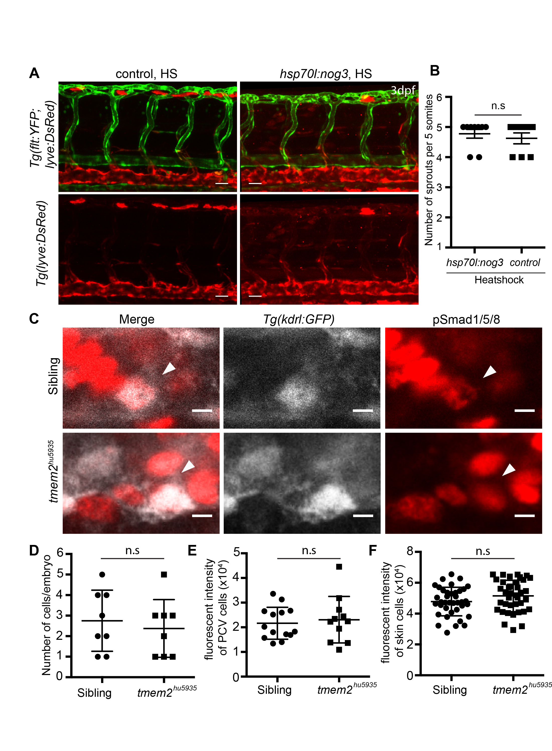
Related to Figure 1: vISV sprouting is correctly patterned following disruption of Bmp signaling and Bmp signaling is unaltered in tmem2 mutants.
A. Lateral view images of 3 dpf Tg(flt1:YFP)/Tg(lyve:DsRed) trunk vasculature in Tg(hsp70l:noggin3) positive and control embryos, heatshocked at 28 hpf for 1 hour, showing normal arterial and venous sprouting under both conditions. Scale bar = 30 μm B. Scatter plots showing quantification of sprouts from the PCV in Tg(hsp70l:noggin3) negative (n=9) versus positive (n=8) embryos (n.s= not significant). C. Whole-mount immunofluorescence staining for pSmad1/5/8 positive cells co-localised with ECs labeled in the Tg(kdrl:eGFP) line in sibling and tmem2 mutant embryos. Scale bar = 5 μm D. Cell counts of EC positive for pSmad1/5/8 show no significant difference between sibling (n=8) and tmem2 mutants (n=8). E. Fluorescence intensity of pSmad1/5/8 positive ECs show no significant difference between sibling (n=14) and tmem2 mutants (n=11). F. Fluorescence intensity of pSmad1/5/8 positive cells was also not significantly different in non-ECs between sibling (n=38) and tmem2 mutants (n=38). Error bars represent ±SD
Reprinted from Developmental Cell, 40, De Angelis, J.E., Lagendijk, A.K., Chen, H., Tromp, A., Bower, N.I., Tunny, K.A., Brooks, A.J., Bakkers, J., Francois, M., Yap, A.S., Simons, C., Wicking, C., Hogan, B.M., Smith, K.A., Tmem2 Regulates Embryonic Vegf Signaling by Controlling Hyaluronic Acid Turnover, 123-136, Copyright (2017) with permission from Elsevier. Full text @ Dev. Cell