
|
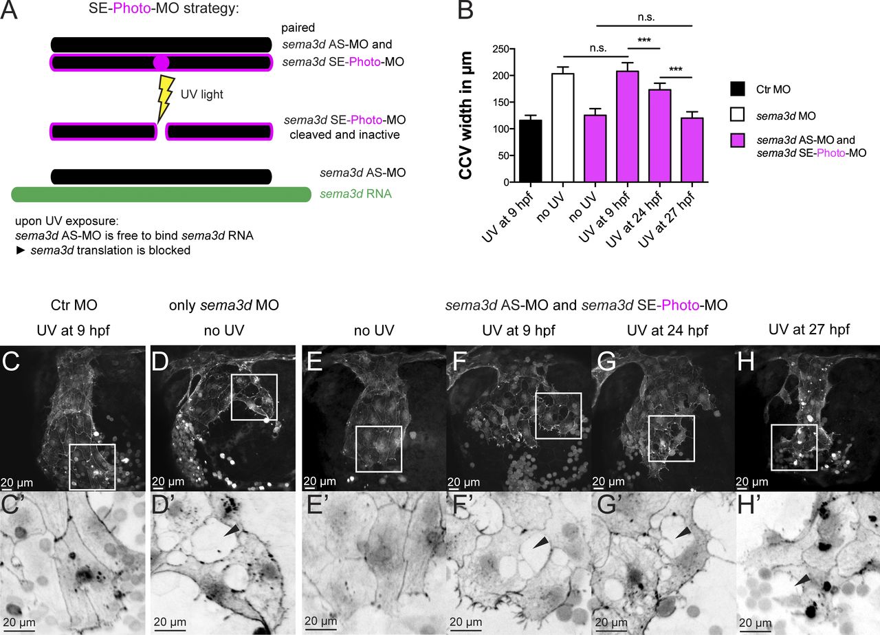
Fig. 4 EC-specific Sema3d acts independently of mesenchymal Sema3d and is required for EC sheet organization and collective migration. Time-specific sema3d knockdown with photo-MOs suggests that EC-specific Sema3d is capable of preserving a cohesive EC sheet. (A) SE-photo-MO strategy for time-specific sema3d knockdown consists of the sema3d AS-MO, paired to a UV light–cleavable sema3d SE-photo-MO (together called AS+SE-photo-MO). UV light induces cleavage of the sema3d SE-photo-MO, enabling the sema3d AS-MO to bind sema3d mRNA and block translation at a chosen time point. (B–H′) Tg(fli1a:lifeactEGFP)mu240 embryos injected with Ctr MO (C), sema3d MO (D), or AS+SE-photo-MO (E–H) were exposed to UV light at the indicated time points and analyzed at 30 hpf. (B) Quantification of CCV front width (each n = 10). ***, P < 0.001; n.s., not significant. Error bars indicate SD. (C and C′) UV exposure has no effect on Ctr MO-injected embryos. (D) sema3d morphants show typical phenotype of widened CCV with lesions in the leading edge (black arrowhead). (E and E′) Without UV induction, the AS+SE-photo-MO–injected embryos resemble Ctr MO. (F and F′) UV activation of AS+SE-photo-MO–injected embryos at 9 hpf results in the sema3d MO phenotype (compare with D and D′). (G and G′) After UV exposure at 24 hpf, AS+SE-photo-MO–injected embryos exhibit an intermediate CCV width (compare with F) but disrupted cell–cell contacts (black arrowhead). (H and H′) AS+SE-photo-MO–injected embryos, exposed to UV light at 27 hpf, exhibit a CCV of normal width (compare with F) but disrupted cell–cell contacts in the front (black arrowhead).