
|
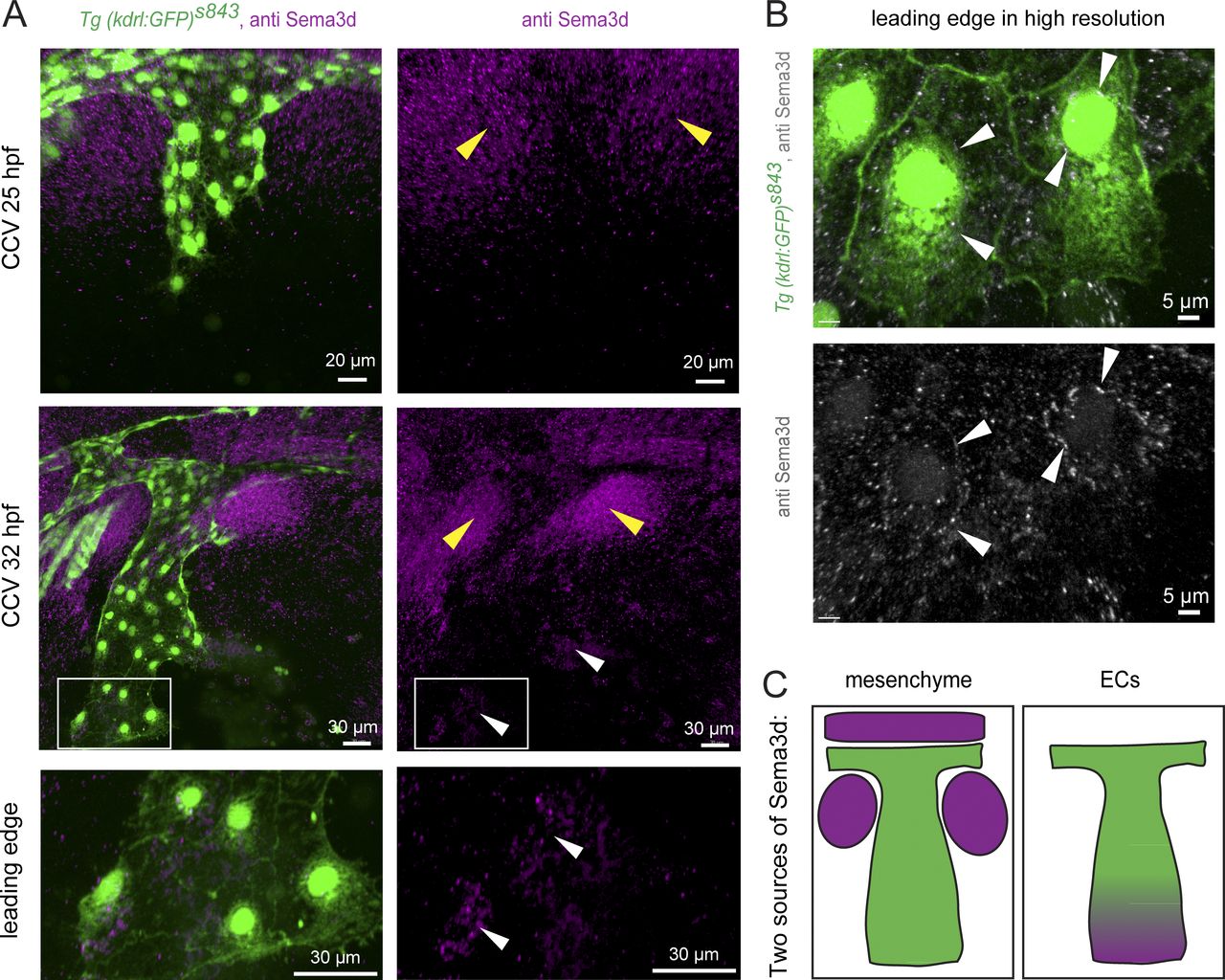
Fig. 2 Sema3d is localized in the mesenchyme next to the CCV and in the ECs of the CCV leading edge. (A) Lateral confocal projections of Tg(kdrl:EGFP)s843 embryos at 25 and 32 hpf stained for Sema3d protein expression. Sema3d is localized in the mesenchyme next to the CCV (yellow arrowheads). At 32 hpf, Sema3d is additionally expressed in the CCV leading edge (white arrowheads). Magnification of the leading edge reveals expression in ECs. (B) Higher-resolution imaging localizes Sema3d expression inside the leading edge ECs at 30 hpf (white arrowheads). (C) Model of the two Sema3d expression domains (magenta, Sema3d; green, ECs).