
|
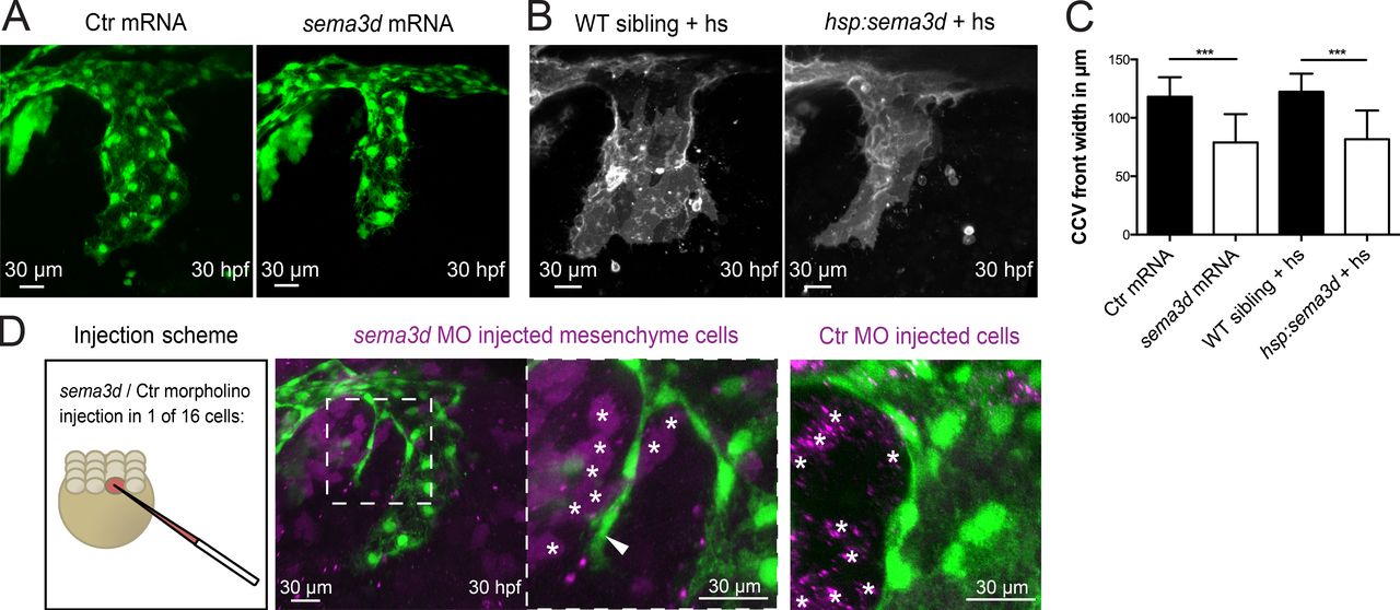
Fig. 3 Mesenchymal Sema3d acts as a repulsive cue for CCV outgrowth. (A–C) Global sema3d overexpression reduces CCV width. (A) sema3d mRNA–injected Tg(kdrl:EGFP)s843 embryos exhibit reduced CCV width at 30 hpf. (B) sema3d expression was induced by consecutive heat shocks (hs) at 16 and 19 hpf (each for 1 h at 39°C) in Tg(hsp70:sema3dGFP), Tg(kdrl:HRAS-mCherry)s896 embryos. sema3d-overexpressing embryos show a thinner CCV at 30 hpf. (C) Quantification of CCV front width (each n = 18). Error bars indicate SD. (D) Mosaic sema3d k.d. through sema3d MO injection into one of 16 cells in Tg(kdrl:EGFP)s843 embryos (magenta, coinjection with DiI for cell tracking). Partial loss of sema3d in the mesenchyme (white asterisks) induced ECs (white arrowhead) to migrate into the mesenchyme (n = 3), indicating that sema3d acts as a repulsive cue in the mesenchyme next to the CCV. Ctr MO-injected mesenchymal cells (white asterisks in right panel) did not induce ectopic EC migration. ***, P < 0.001.