
Fig. 5
- ID
- ZDB-IMAGE-170224-10
- Genes
- Publication
- Hamm et al., 2016 - Sema3d controls collective endothelial cell migration by distinct mechanisms via Nrp1 and PlxnD1
- All Figures
- Figures for Hamm et al., 2016
|
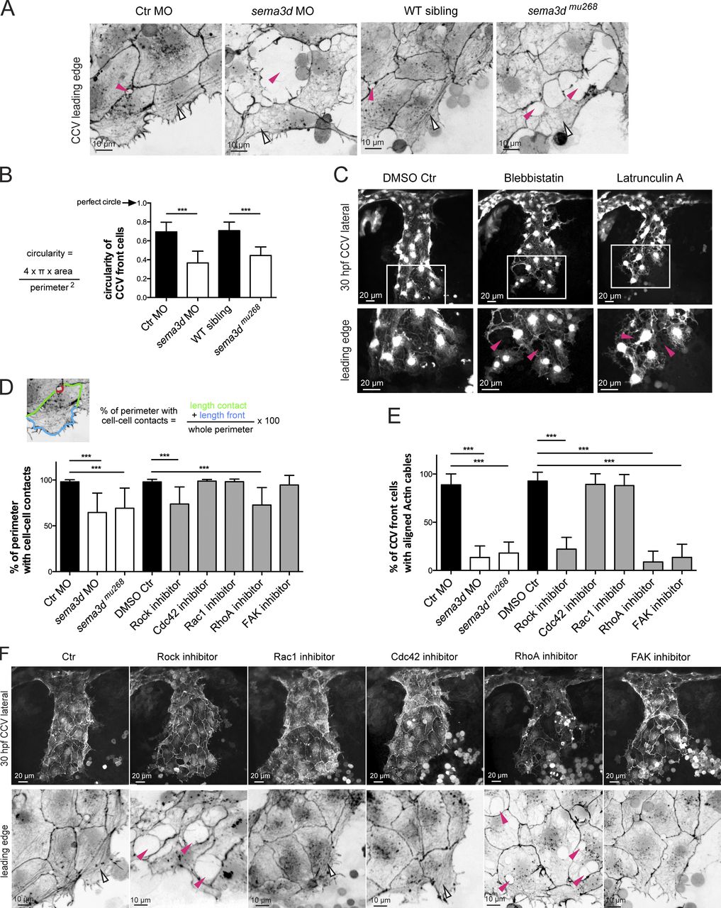
Fig. 5 Sema3d acts on Actin network organization in leading edge ECs controlling Actin cable formation, cell shape, and contacts. (A) The cohesive cell sheet in the CCV leading edge is disrupted and exhibits lesions (pink arrowheads) in sema3d morphants and mutants at 30 hpf. In Ctr embryos, Actin cables are arranged parallel to the leading front; they are absent in Sema3d-deficient embryos (open arrowheads). High-resolution confocal projections of Tg(fli1a:lifeactEGFP)mu240 embryos from Fig. 1 F were color-inverted. (B) Quantification of the circularity of CCV leading edge cells illustrates that loss of sema3d significantly alters cell shape (each n = 24). (C) Inhibition of Myosin/Actin phenocopies sema3d phenotype of disrupted cell–cell contacts (pink arrowheads) but does not lead to a wider CCV. Confocal projections of Tg(kdrl:EGFP)s843 embryos at 30 hpf. (D) The length of cell perimeter with cell–cell contacts of CCV leading edge cells is reduced in sema3d morphants and mutants and after Rock or RhoA inhibition. Inhibition of Rac1, Cdc42, or FAK had no effect on cell–cell contacts (n = 30 for morphants and mutant cells and n = 18 for inhibitor experiments). (E) Quantification of front cells with aligned Actin cables (n = 10 for morphants or mutants and n = 7 for inhibitor experiments). (F) Lateral confocal projections of Tg(fli1a:lifeactEGFP)mu240 embryos at 30 hpf. Leading edge high magnifications were color-inverted. Inhibition of Rock and RhoA induced lesions in the leading edge (pink arrowheads). Parallel-arranged Actin cables (open arrowheads) are missing after inhibition of Rock, RhoA, or FAK. ***, P < 0.001. Error bars indicate SD.