
|
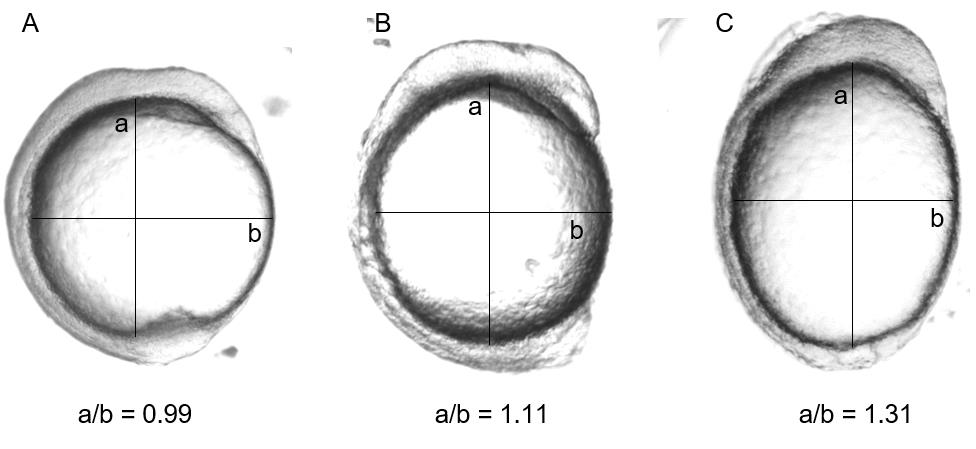
Fig. S1
Measurement of oval shape dimensions of the zebrafish embryo at 11 hpf. Prior to imaging, each embryo was manually positioned so that it was lying laterally. Embryos that died or developed improperly were not considered for measurement. The major (a) and minor (b) axes of the yolk of the embryo were measured on ImageJ, using the Measure feature after drawing a line between the two points demarcating the start and the stop of the axis. The line for measurements were drawn so that the maximum length between the two ends is captured. An example (A) WT-MEK injected embryo, (B) G128V-MEK injected embryo, and (C) F53L-MEK injected embryo are shown.