
Image
|
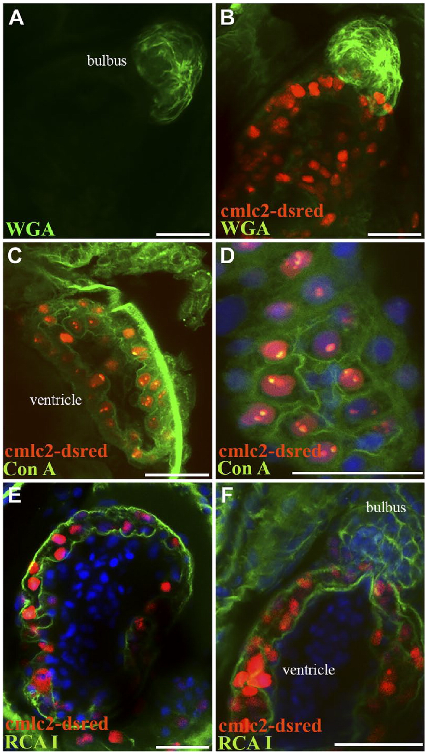
Figure Caption
Fig. 8
Differential lectin binding in developing zebrafish heart. WGA lectin staining of zebrafish heart 72 hr postfertilization shows strong staining of the bulbus mesenchyme (A, B), and little to no staining of the ventricle. Con A stains the endocardium and epicardium of the developing heart (C) as well as cardiac myocyte borders (D). RCA I stains strongly the epicardium and myocardium (E) as well as the developing bulbus (F). Abbreviations: WGA, wheat germ agglutinin; Con A, concanavalin A; RCA I, Ricinus communis agglutinin I. Scale bars: A–F = 10 µm.
Acknowledgments
This image is the copyrighted work of the attributed author or publisher, and
ZFIN has permission only to display this image to its users.
Additional permissions should be obtained from the applicable author or publisher of the image.
Full text @ J. Histochem. Cytochem.