
|
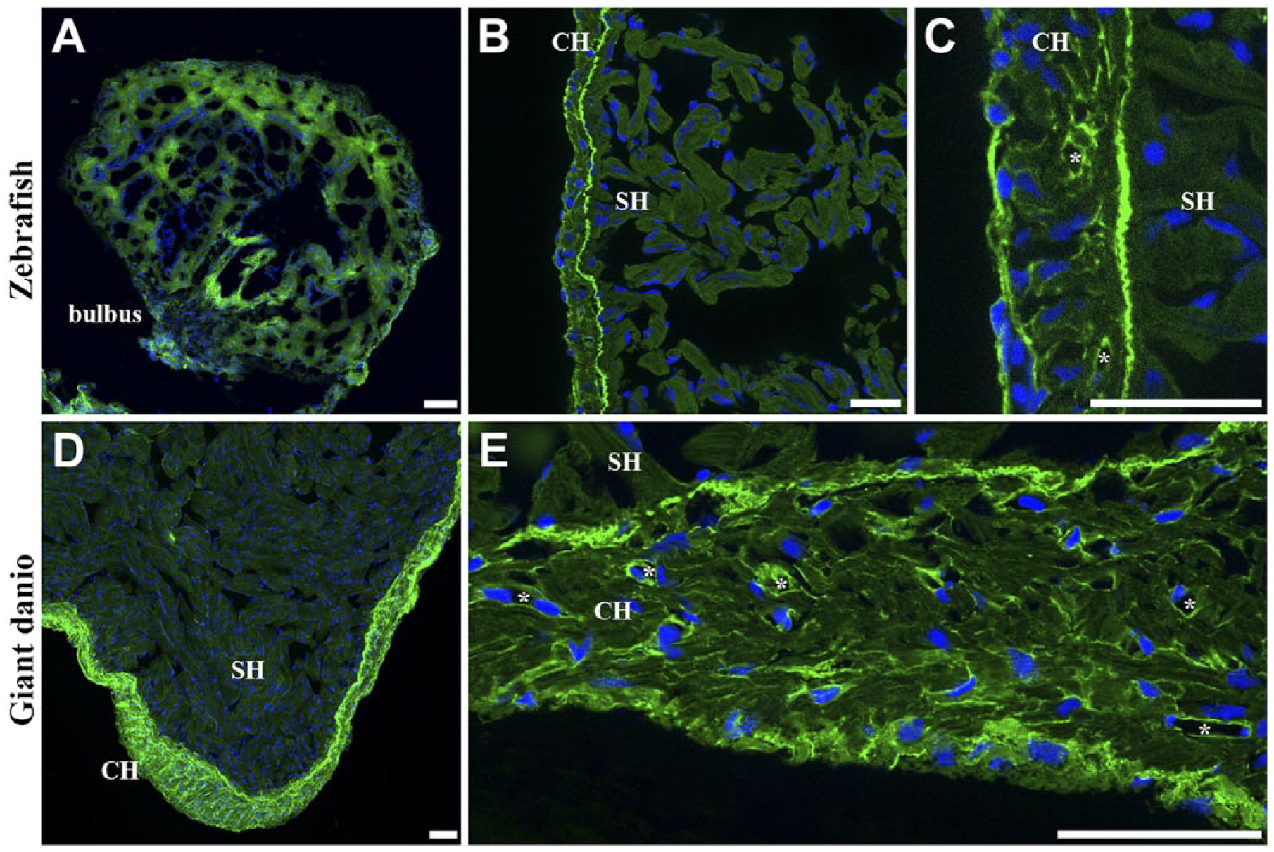
Fig. 6
Differential RCA I lectin binding in giant danio and zebrafish hearts. Low level of RCA I lectin staining was seen in the zebrafish bulbus mesenchyme (A). Strong RCA I lectin binding is seen in the zebrafish CH, and particularly in the junctional region (B). At higher magnification, RCA I staining can be observed in the epicardium, cardiomyocyte borders, and in coronary vessel profiles, albeit at a lower level than the junctional region (C; *, vessel). A similar binding pattern was seen in the giant danio with strong binding in the CH (D) and little to no reactivity in the spongy myocardium. At higher magnification, RCA I is observed to bind strongly in the epicardium, the junctional region, cardiomyocyte borders, and coronary vessel profiles (E; *, vessel). Abbreviations: RCA I, Ricinus communis agglutinin I; SH, spongy heart; CH, compact heart. Scale bars: A–E = 20 µm.