
|
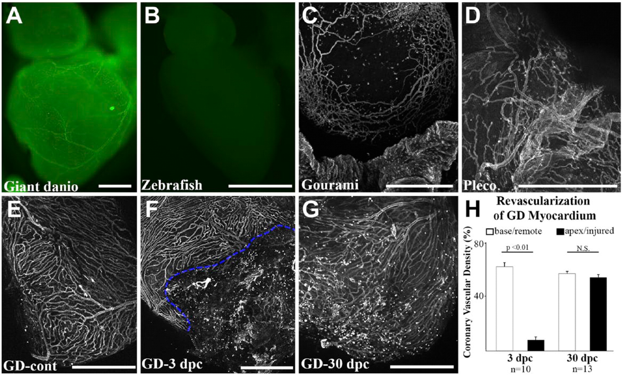
Fig. 5
Whole-mount staining and imaging of fish coronary vasculature. Widefield imaging of Bandeiraea simplicifolia (BS) lectin-fluorescein stained adult giant danio heart where large and small coronary vessels can be visualized (A). Widefield imaging of BS lectin-fluorescein stained zebrafish heart showing absence of BS lectin staining (B). Maximum projection of optical sections showing a vascular network on the gourami bulbus (C) and in the pleco ventricle (D). Maximum projection of optical sections showing a vascular network of uninjured giant danio heart (E) and during regeneration at days 3 (F) and 30 (G) following apical ventricular cautery injury. Dashed line highlights the border zone between injured and non-injured areas of the ventricle. Quantitation of coronary vascularization (H) during regeneration. Scale bars: A–G = 500 µm. GD = giant danio.