
Image
|
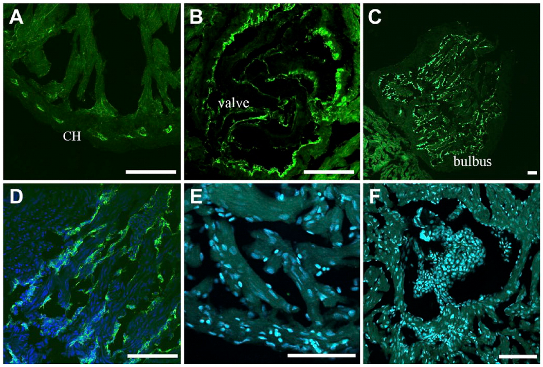
Figure Caption
Fig. 3
Differential BS lectin binding in the giant danio and zebrafish hearts. BS lectin strongly stained the endothelium of the coronary vasculature of the CH, with minimal binding seen in the ventricular endocardium (A). Strong BS lectin binding was seen in the endothelium of the valves (B). BS lectin strongly stained the bulbus (C). Higher magnification show that the staining was localized in the bulbus endothelium folds (D). No BS lectin binding was observed in the zebrafish ventricle (E), including the valves (F). Abbreviations: BS, Bandeiraea simplicifolia; CH, compact heart. Scale bars: A–F = 50 µm.
Acknowledgments
This image is the copyrighted work of the attributed author or publisher, and
ZFIN has permission only to display this image to its users.
Additional permissions should be obtained from the applicable author or publisher of the image.
Full text @ J. Histochem. Cytochem.