
Image
|
Figure Caption
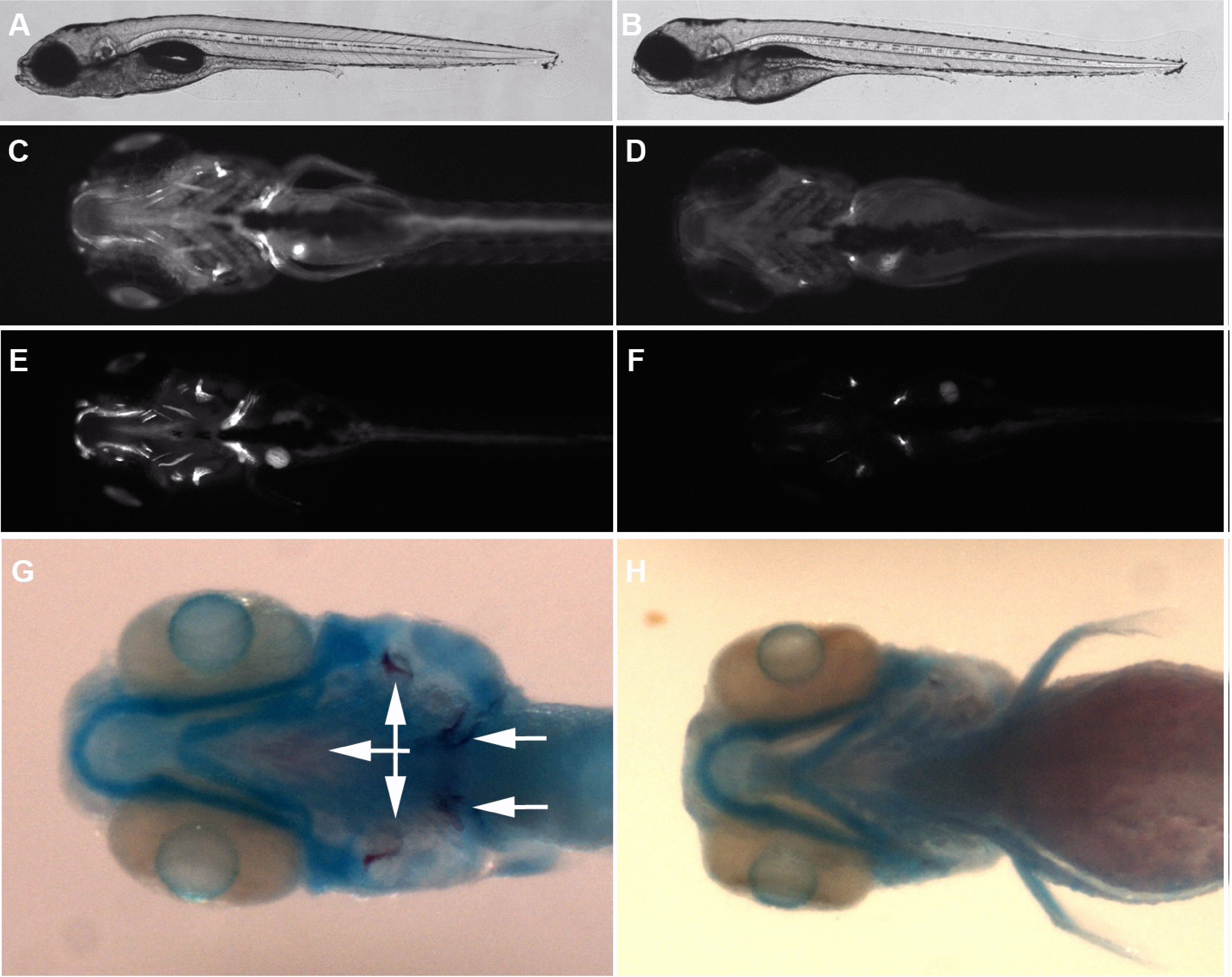
Fig. 2
Bone but not cartilage cells are defective in atp6v1h-deficient zebrafish.
(A-B) shows an image of a 6 days post fertilization (dpf) wild type and mutant embryo with relatively normal gross morphology. (C-F) Day 6 and 8 wild type and mutant embryos showing reduction in bone detected by calcein staining (ventral view, bright fluorescent stains indicate calcified bone). In (G-H), double staining for cartilage and bone demonstrate reduction of mineralized bone (dark purple, arrows) but not cartilage (blue). A, C, E and G are wild type. B, D, F, and H are mutant.
Figure Data
Acknowledgments
This image is the copyrighted work of the attributed author or publisher, and
ZFIN has permission only to display this image to its users.
Additional permissions should be obtained from the applicable author or publisher of the image.
Full text @ PLoS Genet.