
|
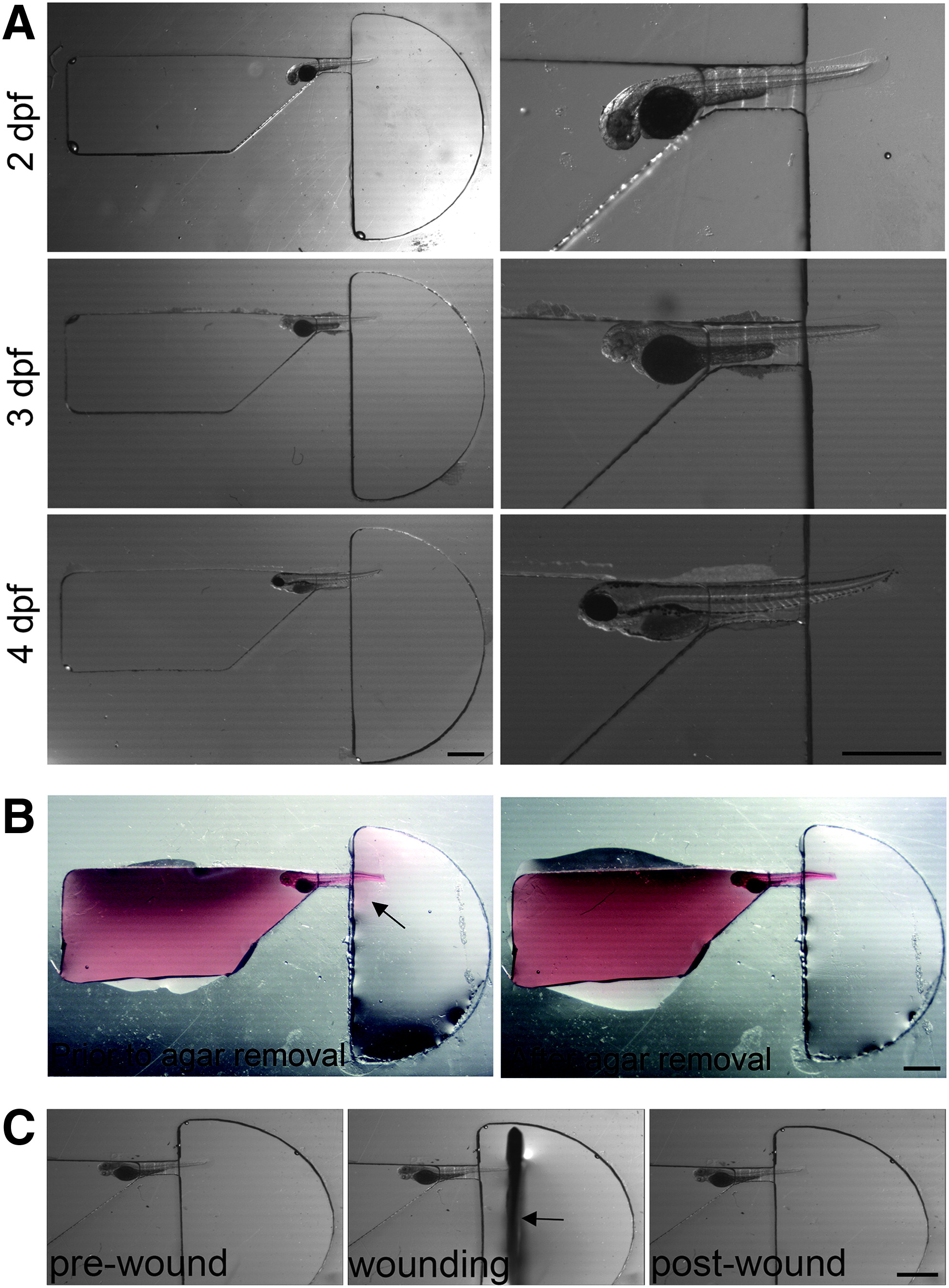
Fig. 2
Positioning of larva in channel. (A) Dissecting microscope images showing 2, 3, and 4 dpf fixed larvae loaded into zWEDGI channel, with enlargement to show detail. (B) A minimal amount of agarose, labeled with rhodamine 6G and filling the loading chamber, exits from restraining tunnel into the wounding chamber, as detected by the pink halo (arrow). Residual agarose can be removed using a syringe needle with minimal impact on the tail, leaving the tail unrestrained in buffer in the wounding chamber. (C) Semicircular wounding chamber is designed to accommodate scalpel blade (arrow) for caudal fin transection. Scale bar = 1 mm. dpf, days postfertilization.