
|
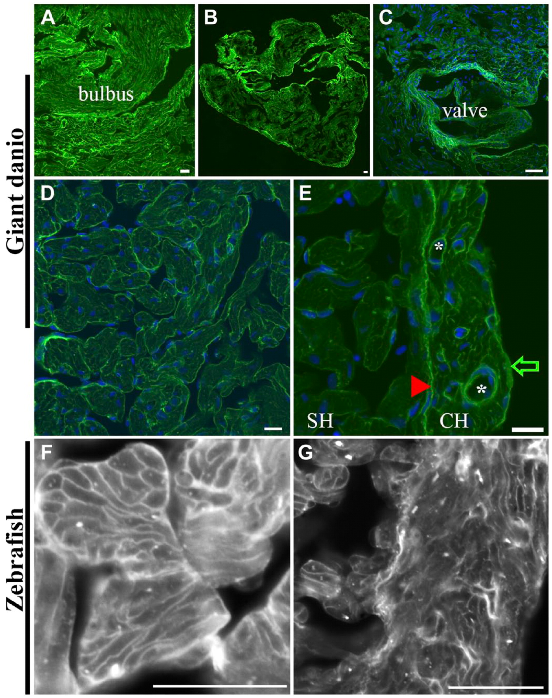
Fig. 1
Differential Con A lectin binding in the giant danio and zebrafish hearts. Con A-fluorescein lectin binding (green) is observed throughout the giant danio heart with the strongest staining seen on the bulbus (A), the CH (B), and the valves (C; nuclei, blue). At higher magnification, Con A binds to the trabecular bundles in the spongy myocardium and to cardiomyocyte membranes (D). Con A binding is comparatively stronger in the CH compared with that in the SH (E). Within the compact myocardium, Con A binding is particularly strong in the epicardium (arrow) and the coronary vasculature (*). The junctional region (arrowhead) is equally stained, forming a boundary line between the CH and SH, with small regions of discontinuity at regular intervals. A similar pattern is seen in the zebrafish heart with Con A staining cardiomyocyte borders of the SH (F) and CH (G). Abbreviations: Con A, concanavalin A; SH, spongy heart; CH, compact heart. Scale bars: A–G = 10 µm.