
|
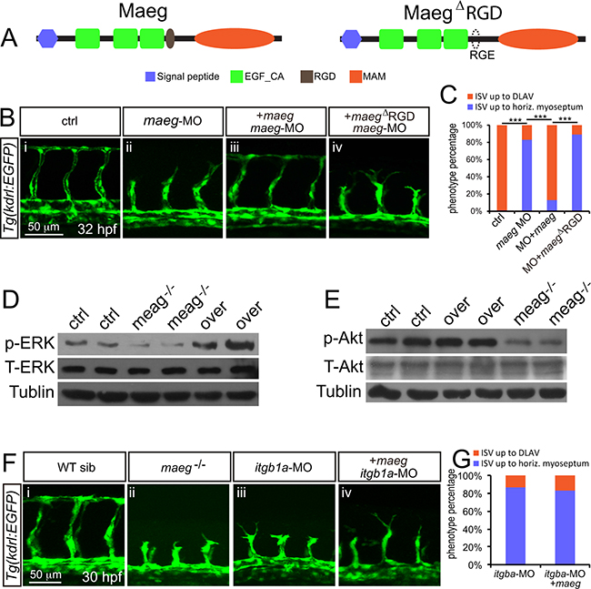
Fig. 6
Maeg regulates angiogenesis dependent on RGD domain. A. The diagram of Maeg protein with RGD domain and Maeg protein with RGD domain mutated to RGE domain (MaegΔRGD). B. Confocal images of ISVs in control embryos, maeg morphants, maeg morphants treated with DAPT, and maeg morphants coinjected with dll4 MO using Tg(kdrl:EGFP) transgenic embryos. C. Percentage of embryos with ISV defect in each group. χ2 test; ***, P<0.001. D, E. Phosphorylation levels of ERK and Akt determined by Western blot analysis in maeg loss- and gain-of-function embryos. F. Confocal images of ISVs in control embryos, maeg mutants, itgb1a morphants, and itgb1a morphants coinjected with maeg mRNA using Tg(kdrl:EGFP) transgenic embryos. G. Percentage of embryos with ISV defect in each group.