
|
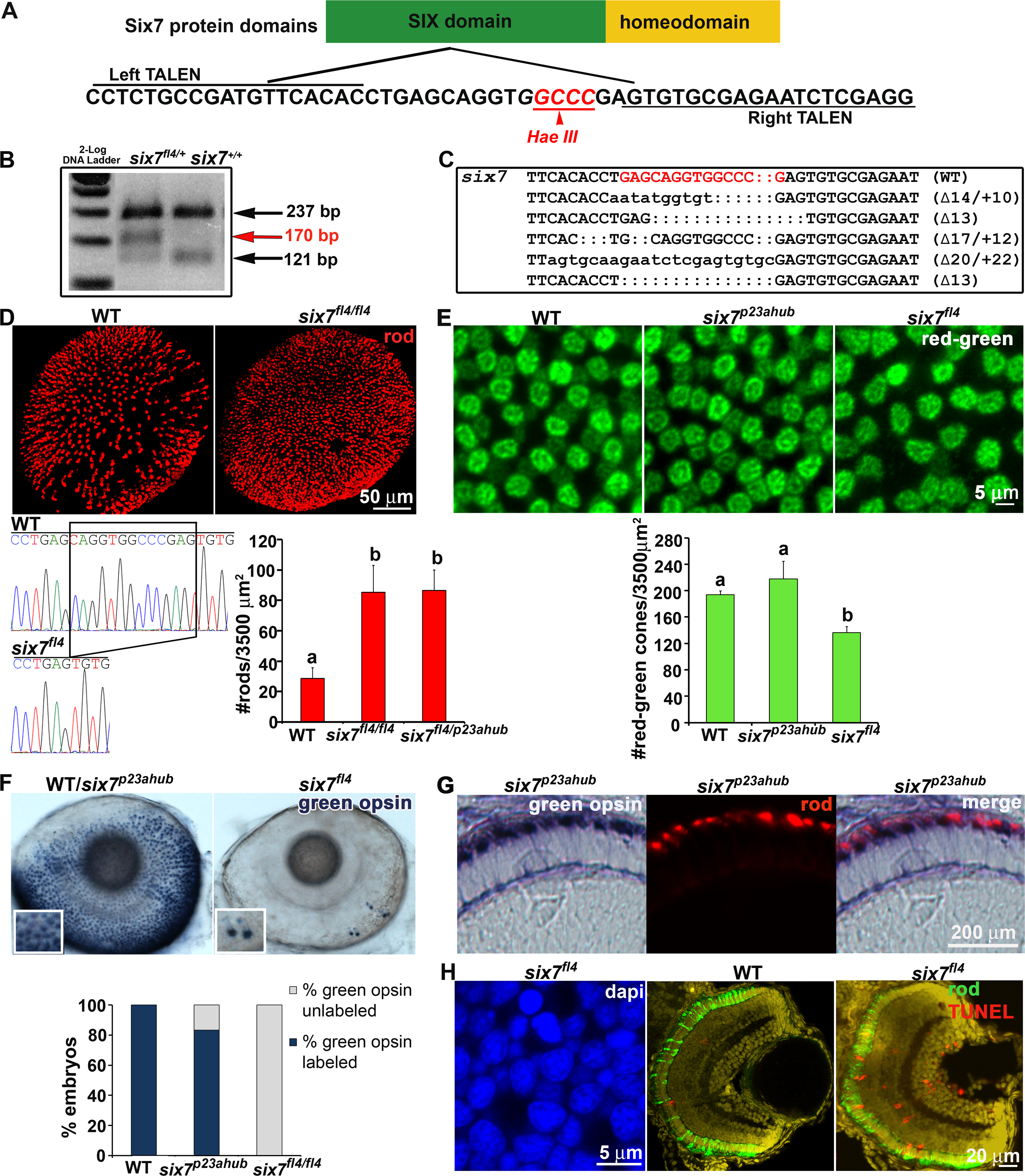
Fig. 4
TALENs-mediated knockout of six7 locus recapitulates ljrp23ahub phenotype.
(A) Schematic representation of the Six7 protein domains. six7-TALENs target site and the HaeIII restriction enzyme site are highlighted. The left and right monomer binding sites are underlined. (B) RFLP of six7 locus by HaeIII. A new 170 bp DNA-fragment is detected in six7fl4 carriers compared with controls. (C) Sequence analysis of the six7 target region shows recovery of multiple indel alleles of six7. The target region is highlighted in red. Dots represent deletions and lower case letters indicate insertions. The six7 sequence from WT is shown as a comparison. (D) Confocal immunofluorescent images labeled for rods (red) from WT and six7fl4 knockout mutants at 4 dpf. six7fl4 knockout mutants phenocopy ljrp23ahub mutants. Sequencing chromatograms of WT and six7fl4 mutants illustrated the c.217_229del CAGGTGGCCCGAG (del13) six7 mutation in homozygous zebrafish. The bar graph shows the average number of rods counted in 1–3 different areas per retina (WT (n = 8), six7fl4 (n = 7), six7 fl4/p23ahub (n = 4); One-way ANOVA with Tukey’s post-hoc test. a vs b, p<0.0001. (E) Tangential views of confocal immunofluorescent images labeled for red-sensitive (brighter signal) and green-sensitive (dimmer signal) cones from WT, six7p23ahub, and six7fl4 retinas at 4 dpf. Graph showing the average number of red/green-sensitive cones per unit area of WT (n = 3), six7p23ahub (n = 4), and six7fl4 (n = 3); (ns p> 0.05; a vs b p ≤ 0.05; significant difference one-way ANOVA with Tukey’s post-hoc test). (F) Whole mount in situ hybridization for RH2 probe (green-sensitive cone opsin) in WT (n = 30), six7p23ahub (n = 30) and six7fl4 embryos (n = 30) at 4 dpf. WT and six7p23ahub embryos often showed the same pattern and number of green-sensitive opsin cone labeling (Dorsal is up and nasal to the left), while six7fl4 knockout showed no labeling or few cells labeling for green-sensitive cone opsins (inset). Graph showing the percentage of unlabeled and labeled embryos. Notice that 17% of the six7p23ahub embryos were un-labeled for green-sensitive cone opsin. (G) Retinal cryosections of in situ hybridization for green-sensitive cone opsin in six7p23ahub (n = 5) embryos immunolabeled with 1D1 (rods). Rods and green-sensitive cone opsin probes labeled different cells in the ONL of six7p23ahub embryos. (H) Evidence of cell death. Flat mount confocal image of nuclei counterstained with DAPI and retinal cryosections from WT and six7fl4 animals at 4dpf co-labeled for TUNEL (red) and rods (4C12, green). six7fl4 mutants (n = 6, 1–2 sections/retina) showed an increase in apoptotic cells, especially in the ONL compared with WT (n = 3, 1–2 sections/retina), (arrows pointing to apoptotic cells in the ONL).