
|
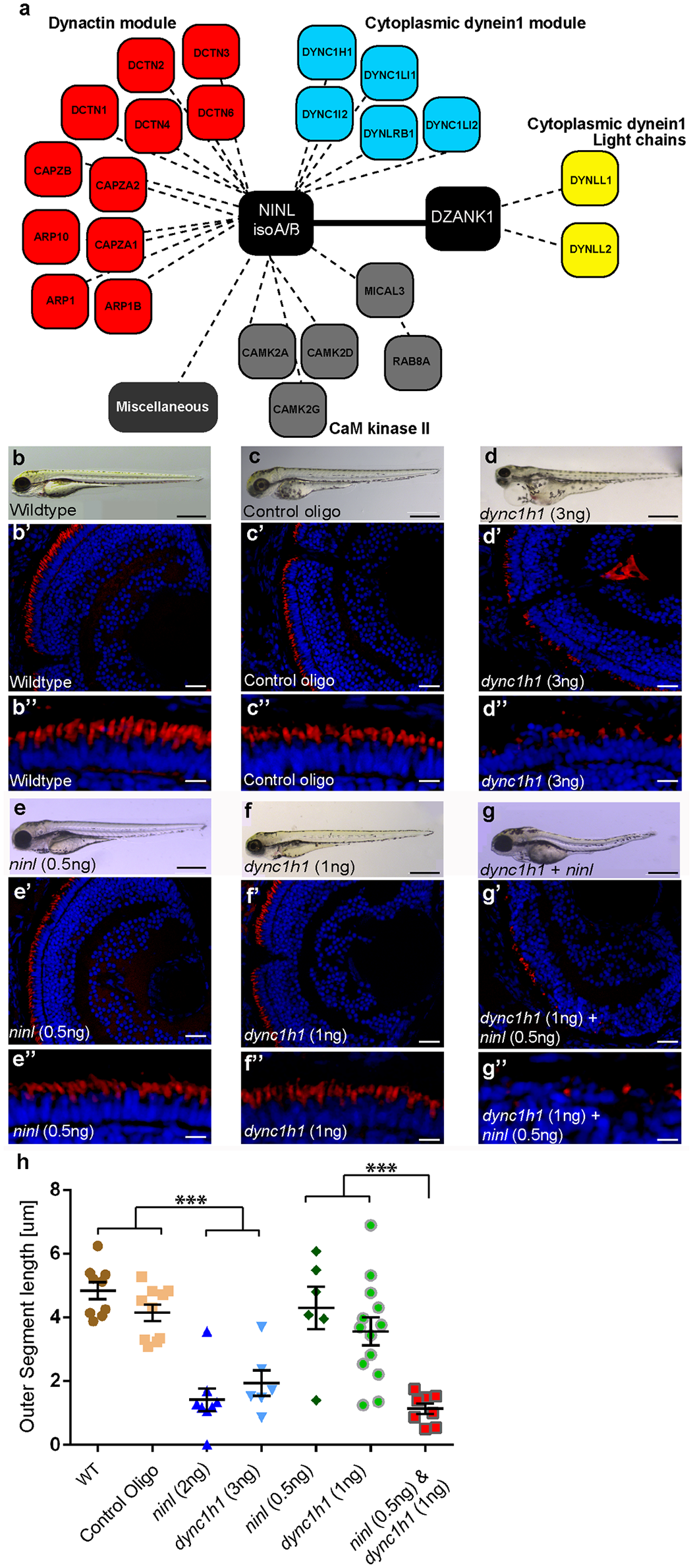
Fig. 5
NINL and DZANK1 associate specifically with complementary subunits of the cytoplasmic dynein 1 motor complex.
(a) Strep-SILAC and TAP experiments show that DZANK1 interacts specifically with DYNLL1 and DYNLL2 (yellow). NINL associates with components of the dynactin complex (red) and with most components of the dynein 1 motor complex (blue), except for DYNLL1 and DYNLL2. The solid line between NINL and DZANK1 symbolizes a direct interaction, whereas the dashed lines stand for interactions determined by immune and affinity purifications. (b-g'') Genetic interaction between ninl and dync1h1. MO-injection with a high concentration of dync1h1 (3 ng/nl) shows larvae with pericardial edema, small eyes and short OS (d-d''). MO-injection of sub-effective concentrations of ninl (0.5 ng/nl) (e-e'') or dync1h1 (1 ng/nl) (f-f'') generates larvae with a normal phenotype and normal OS length, compared to wildtype larvae (b-b'') and control MO-injected larvae (c-c''). The combination of sub-effective MO concentrations of ninl (0.5 ng/nl) and dync1h1 (1 ng/nl) results in short larvae and virtual absence of outer segments (g-g''). All larvae are 4 dpf. Panels b'-d'' and e'-g'' are retinal cryo-sections stained with bodipy (red) to highlight the outer segments and DAPI to stain the nuclei. (h) Quantification of photoreceptor outer segment length revealed a significantly decreased length of outer segments in dync1h1 (3ng/nl) and dync1h1/ninl double morphants as compared to controls (*** P<0.0001; two-tailed, unpaired Student's t-test). Scale bars represent 500 μm (b-g), 50 μm (b'-g') and 15 μm (b''-g'').