
|
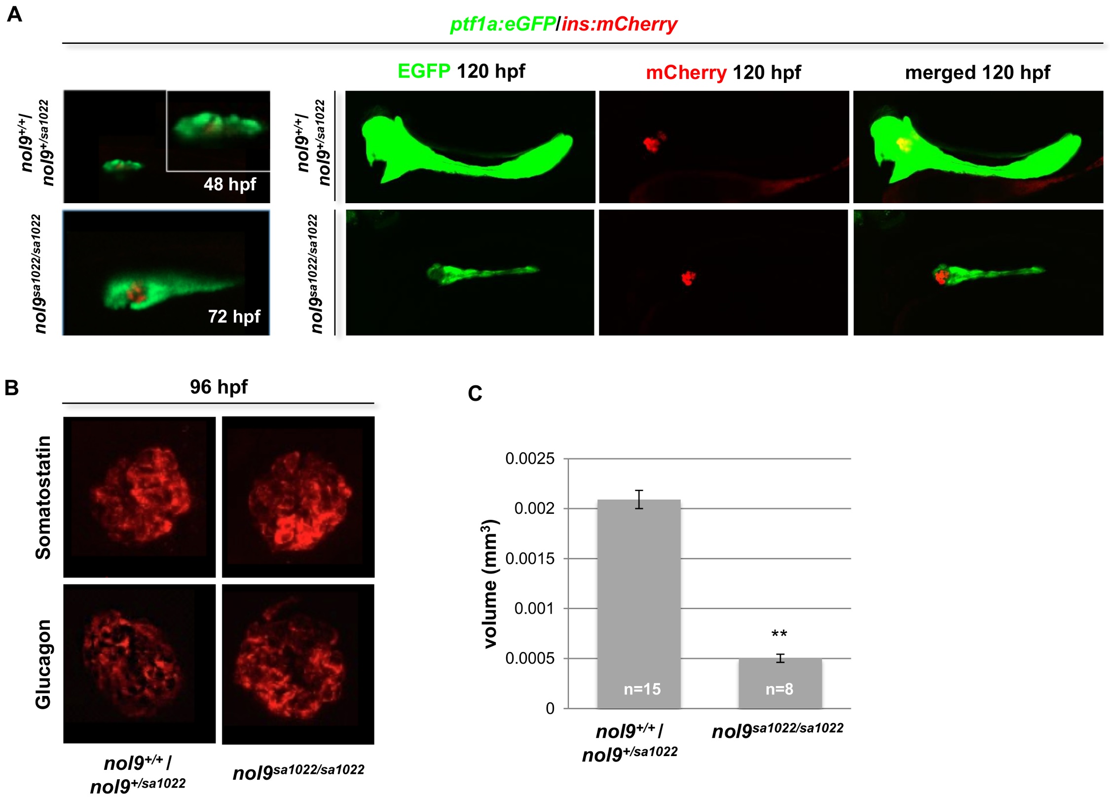
Fig. S4
nol9sa1022/sa1022 mutants have a defect in development of the exocrine but not the endocrine pancreas.
(A) Representative confocal images of the pancreas of Tg(ptf1a:EGFP;ins:mCherry) fish at 48-, 72- and 120 hpf. No difference was observed between nol9sa1022/sa1022 and wt siblings at 48- and 72 hpf. At 120 hpf the area of the ptf1a+ exocrine pancreas (green) was decreased in nol9sa1022/sa1022 larvae, while the area of ins+ endocrine pancreas (red) was comparable in nol9sa1022/sa1022 mutants and wt siblings. (B) Confocal images of the endocrine pancreas of larvae subjected to immunohistochemistry against Somatostatin and Glucagon at 96 hpf. The intensity and area covered by the signal was comparable in nol9sa1022/sa1022 larvae and wt siblings for both antibodies. (C) Average volume of the ptf1a-positive exocrine pancreas in nol9sa1022/sa1022 (n = 8) and wt (n = 15) larvae at 120 hpf. Data are represented as the mean +/- SEM, Student’s t-test, **, p<0.01. All images are oriented with anterior to the right and dorsal to the top.