
Fig. S1
- ID
- ZDB-IMAGE-170208-17
- Publication
- Bielczyk-Maczyńska et al., 2015 - The Ribosome Biogenesis Protein Nol9 Is Essential for Definitive Hematopoiesis and Pancreas Morphogenesis in Zebrafish
- All Figures
- Figures for Bielczyk-Maczyńska et al., 2015
|
Fig. S1
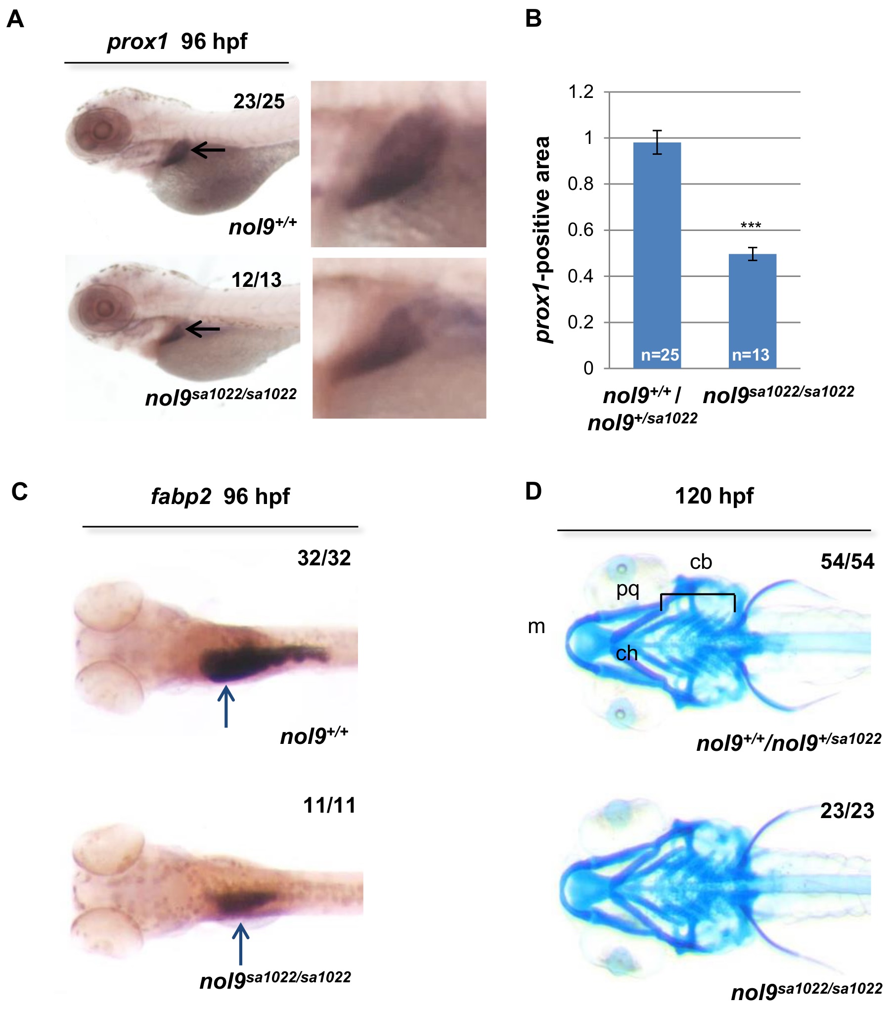
nol9 mutation affects the development of liver and intestine but not of jaw cartilage.
(A) Representative pictures of 96 hpf larvae stained by WISH against the liver marker prox1 (black arrow). Magnified images of the liver are shown. (B) Quantification of the prox1 in situ hybridization data. The average prox1-positive area is decreased in nol9sa1022/sa1022 mutants (n = 13) compared to wild-type siblings (n = 25). Data are represented as the average +/- SEM. Two-tailed Student’s t test, ***, p<0.001. (C) Representative images of 96 hpf larvae stained by WISH against the intestinal marker fabp2a. The expression of fabp2a (blue arrow) is decreased in nol9sa1022/sa1022 (n = 11) compared to wild-type siblings (n = 32). (D) Alcian blue staining, showing normal formation of the jaw cartilage elements Meckel’s (m), palatoquadrate (pq), ceratohyal (ch) and ceratobranchial (cb) in both nol9sa1022/sa1022 mutants (n = 23) and wt siblings (n = 54) at 120 hpf. Ventral view with anterior to the left.