
|
Fig. S1
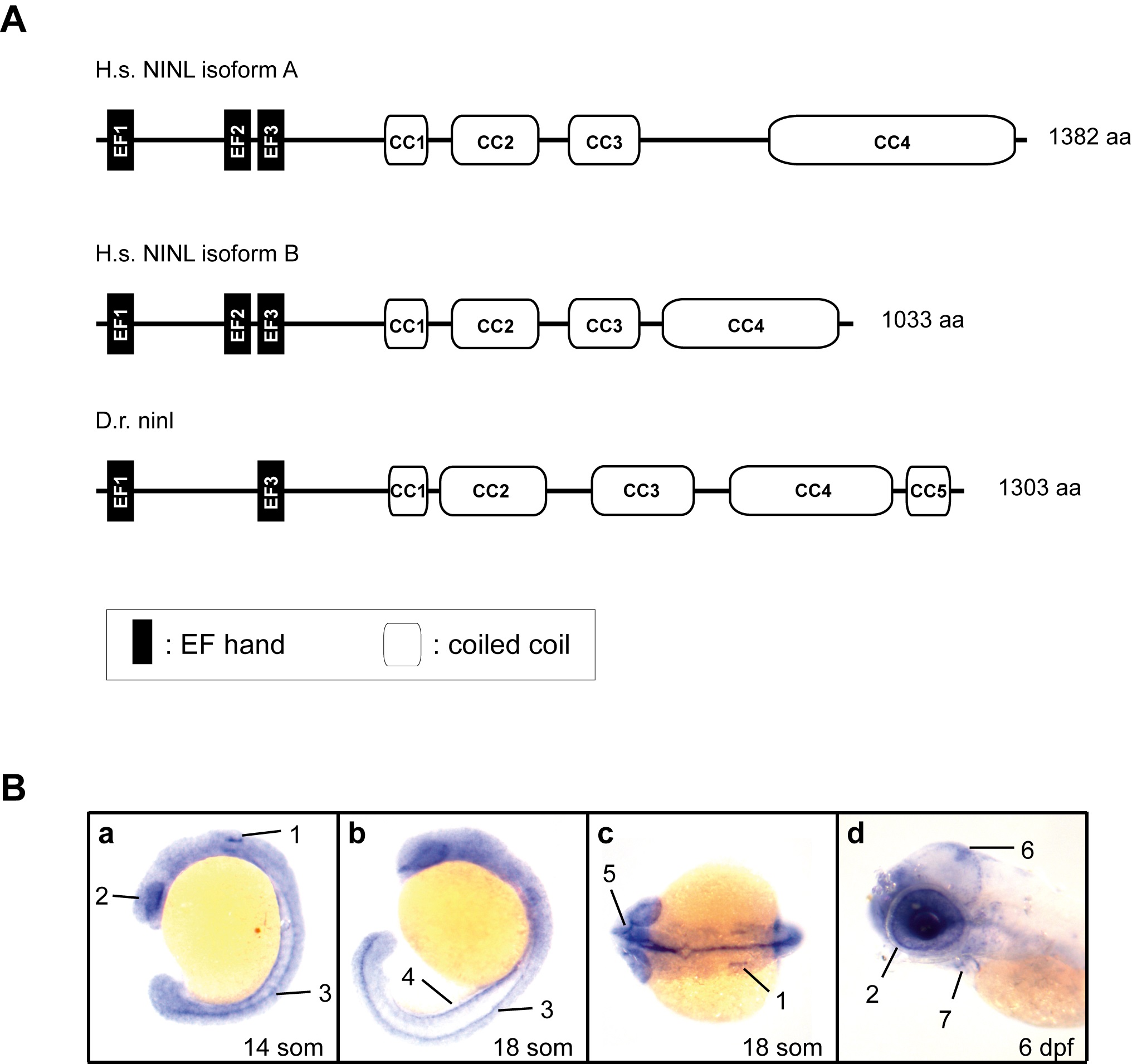
Cloning and characterization of zebrafish ninl.
(A) Schematic representation of the protein structure of human NINLisoA and NINLisoB (H.s. NINLisoA and H.s. NINLisoB) and zebrafish ninl (D.r. ninl) as predicted by using the Pfam homepage (http://pfam.xfam.org). (B) Ninl expression during zebrafish development by whole mount RNA in situ hybridization. Specific expression was found in the following structures as indicated by numbers and arrows: (a) 14 somite stage: otic placode (1); developing eye (2); neural tube (spinal cord) (3); (b) 18 somite stage: neural tube (3); pronephros (4); (c) 18 somite stage: inner ear (1); optic nerve (5). (d) At 6 dpf, expression was observed in the tectum (6), the heart (7) and in the eye (2), predominantly in the photoreceptor cell layer. H.s.: homo sapiens; D.r.: danio rerio; CC: coiled-coil; IF: intermediate filament domain; som: somites; dpf: days post-fertilization.