
Fig. 2
- ID
- ZDB-IMAGE-170207-1
- Genes
- Antibodies
- Source
- Figures for Bachmann-Gagescu et al., 2015
|
Fig. 2
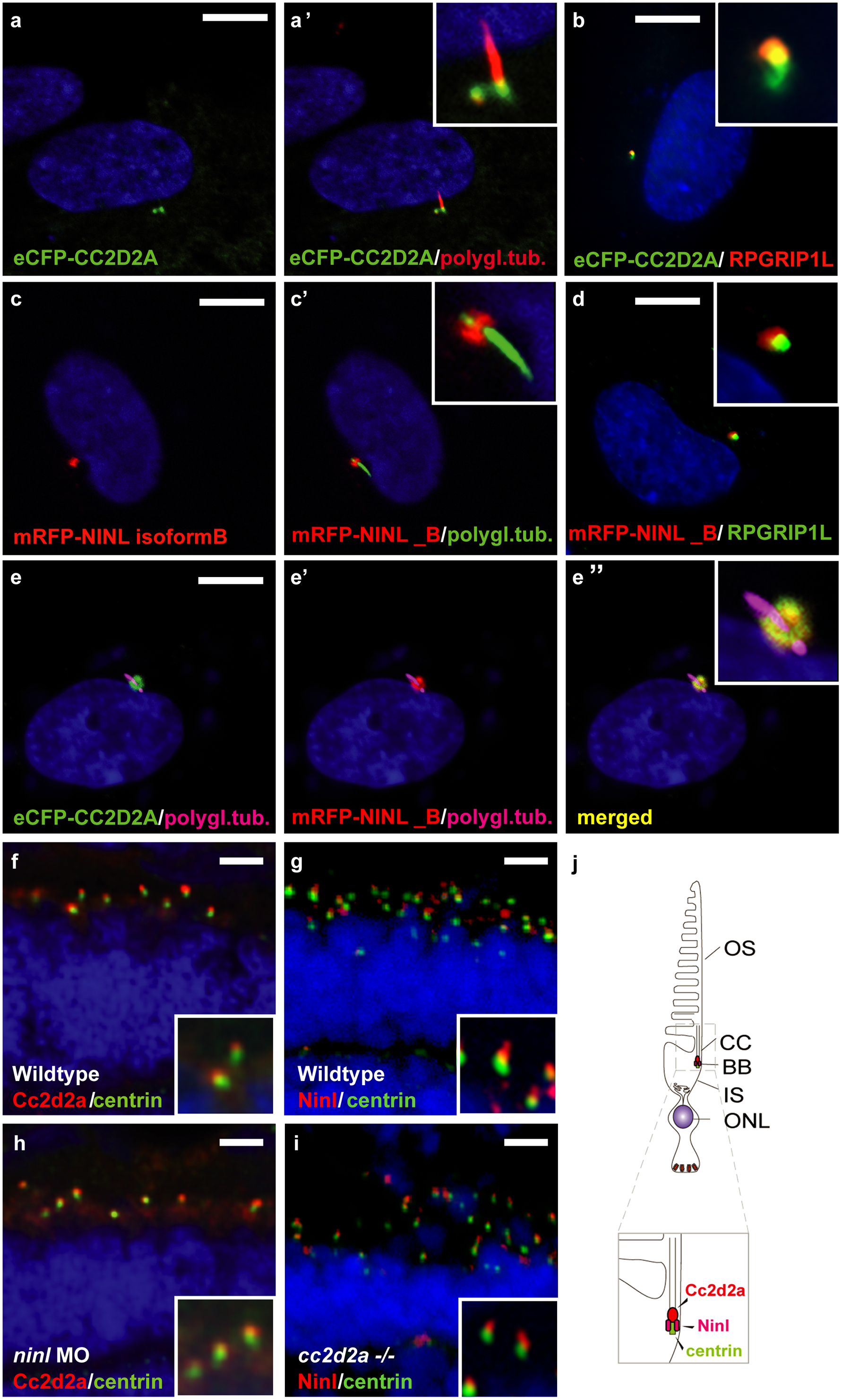
CC2D2A and NINL co-localize at the ciliary base in hTERT-RPE1 cells and in zebrafish retina.
(a, a' and inset) When expressed alone, eCFP-tagged CC2D2A (green signal) localizes to the ciliary base (basal body, accessory centriole). The cilium is marked by anti-polyglutamylated tubulin (red signal, a' and inset). eCFP-tagged CC2D2A (green signal; b) also (partly) localizes to the ciliary transition zone, which was visualized using anti-RPGRIP1L as a marker (red signal; b). (c, c' and inset) mRFP-tagged NINL isoform B was localized at the ciliary base (cilium in green, c' and inset). (d and inset) mRFP-tagged NINL isoform B (red signal) localizes adjacent to the ciliary transition zone (anti-RPGRIP1L; green signal). (e-e” and inset) Co-expression of mRFP-tagged NINL isoform B (red signal) and eCFP-tagged CC2D2A showed co-localization of both proteins around the ciliary base (yellow signal). (f) In wild-type larval zebrafish retina (4 dpf), Cc2d2a marked by anti-Cc2d2a antibodies (red signal) is localized apically to the photoreceptor basal body (marked by anti-centrin antibodies, green signal). (g) Ninl, stained with anti-Ninl antibodies, (red signal) is localized at the zebrafish photoreceptor ciliary base, partially overlapping with and apical to the green centrin signal. (h) Cc2d2a localization is unaffected by ninl knockdown and (i) Ninl localization is normal in cc2d2a-/- larvae. (j) Schematic representation of the localization of Ninl and Cc2d2a in zebrafish photoreceptor cells. (f-i) are immunostainings on cryosections from 4 dpf larvae. Nuclei were stained with DAPI (blue signal) in all panels. Scale bars are 10 μm in a-e, and 4 μm in f-i.