
|
Fig. 4
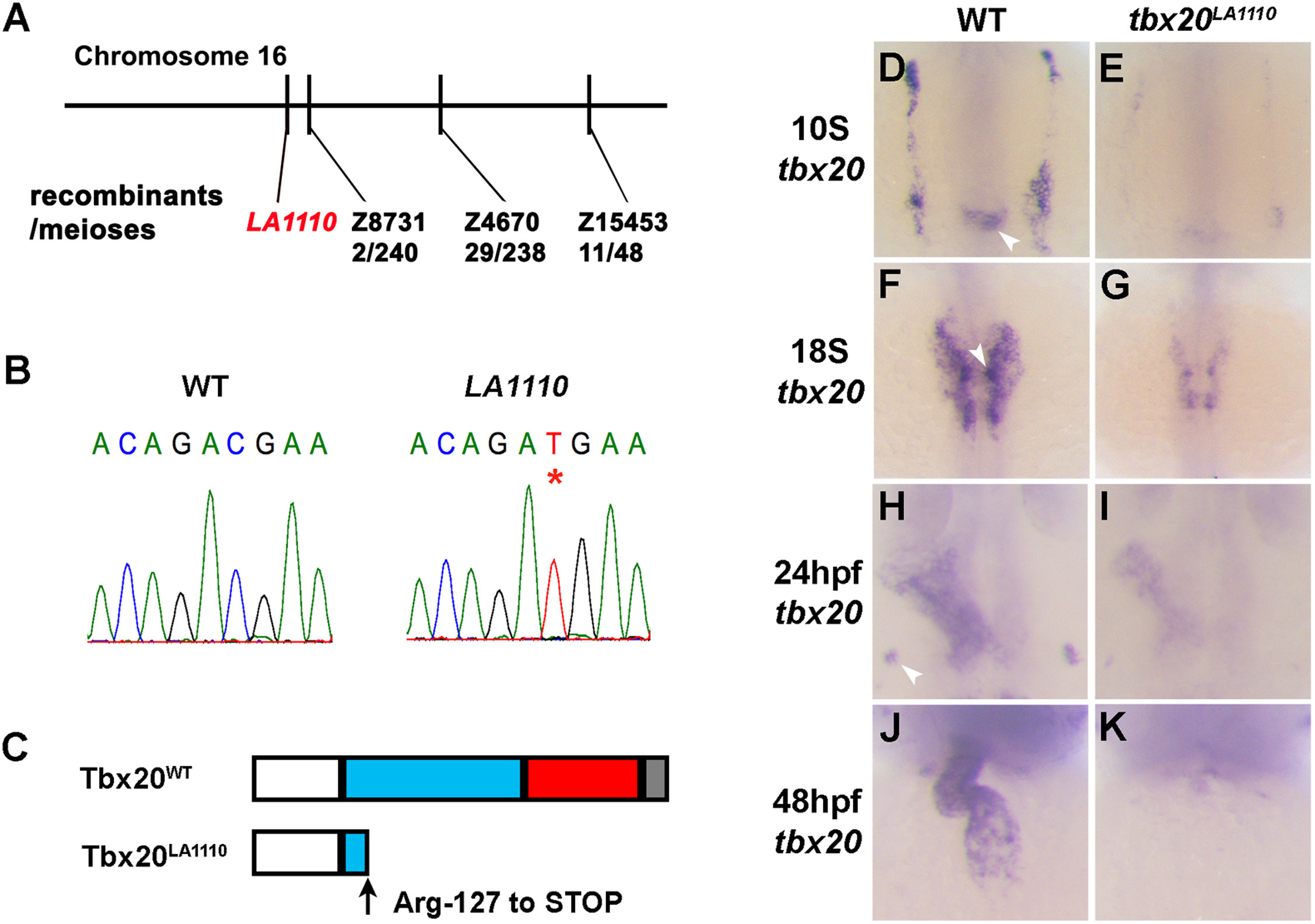
Positional cloning of the tbx20LA1110 mutant. (A) Diagram showing the location of the LA1110 locus with respect to markers on Chromosome 16. (B) Sequencing of the tbx20 locus in wild type and LA1110 mutant embryos identified a C to T transition, resulting in a premature stop codon (*). (C) Schematic diagram of the Tbx20WT and Tbx20LA1110 proteins. Zebrafish Tbx20WT contains an N-terminal domain (white), a T-box domain (blue), and predicted transactivation (red) and transrepression (grey) domains in the C-terminal portion of the protein. The Tbx20LA1110 protein is truncated within the T-box DNA binding domain. (D-K) Expression of tbx20 in wild type and LA1110 embryos showing the anterior dorsal view (D-I) and frontal view (J, K). In wild type embryos, expression of tbx20 is visible as bilateral strips in the cardiac primordia within the ALPM at the 10-somite stage (D). These strips of cells migrate toward and fuse at the midline (F). By 24 hpf, the entire heart tube displays expression of tbx20 (H, J). Additional tbx20 expression is present within the hindbrain at the 10-somite stage (arrowhead in D), hindbrain branchiomotor neurons at the 18-somite (F) and the primordia of the pituitary gland at 24 hpf (arrowhead in H). (E, G, I and K) In tbx20LA1110 mutant embryos, tbx20 transcripts are dramatically reduced.
Reprinted from Developmental Biology, 421(2), Lu, F., Langenbacher, A., Chen, J.N., Tbx20 drives cardiac progenitor formation and cardiomyocyte proliferation in zebrafish, 139-148, Copyright (2017) with permission from Elsevier. Full text @ Dev. Biol.