
Image
|
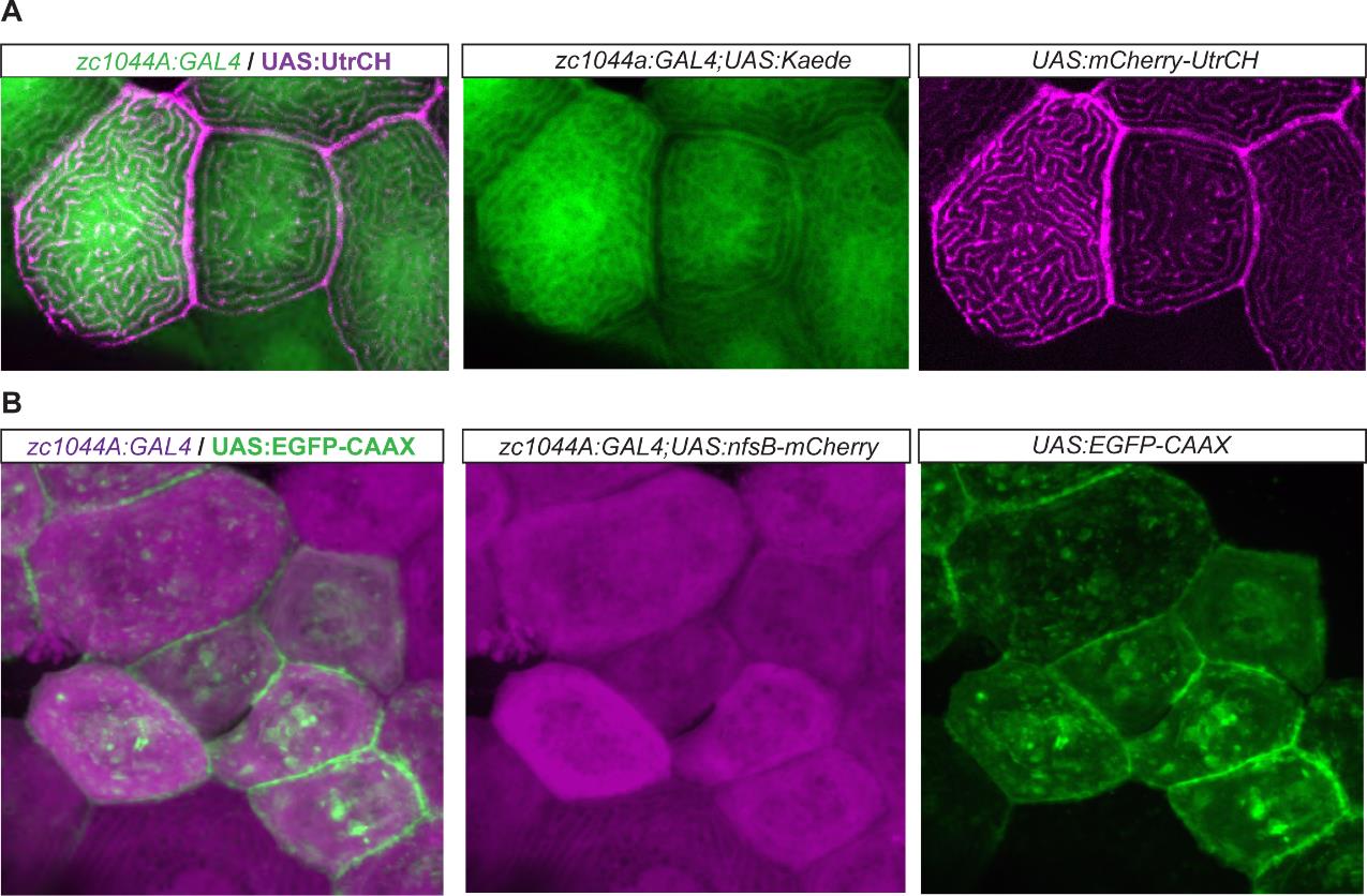
Figure Caption
Fig. S4
Epithelial enhancer trap line driving expression of UAS cytoskeletal fluorescent reporter lines. Expression of the UAS cytoskeletal fluorescent reporters by the Periderm-GET line reveals localization of F-actin (A) and membrane bound fluorescent expression (B), respectively, within the developing periderm (48hpf). (A). Expression of UAS:mCherry-UtrCH (magenta) in the Et(Gal4-VP16)zc1044A; Tg(UAS-E1b:Kaede)s1999t (green) line; and (B) UAS:EGFP-CAAX (green) in Et(Gal4- VP16)zc1044A;Tg(UAS-E1b:nsfB-mCherry) (magenta) line.
Acknowledgments
This image is the copyrighted work of the attributed author or publisher, and
ZFIN has permission only to display this image to its users.
Additional permissions should be obtained from the applicable author or publisher of the image.
Full text @ J. Cell Sci.