
|
Fig. 1
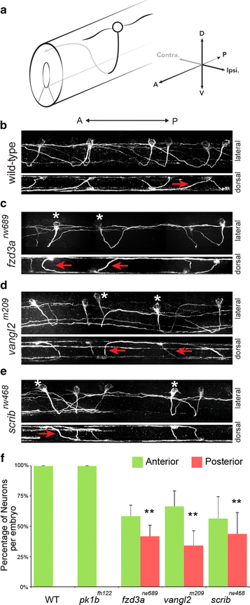
PCP genes are required for anterior pathfinding of CoPA axons after midline crossing. a Illustration of a CoPA neuron in the spinal cord. Black line represents ipsilateral ventral projection, Gray line indicates dorso-anterior trajectory after midline crossing. CoPA neurons have two major dendrites, one projecting anteriorly, and the other projecting posteriorly, b confocal micrographs of 3A10 immunofluorescence shows the pathfinding of CoPA axons in multiple segments of the spinal cord in wild-type (WT) embryos. In the lateral view (upper panel), all CoPA axons project dorso-anteriorly after midline crossing. Anterior is to the left, dorsal is up. In the lower panel, a dorsal view of the same spinal cord shows the midline crossing trajectory of CoPA axons (red arrows), c–e in fzd3 rw689 , vangl2 m209 , and scrib rw468 mutant embryos, approximately half of CoPA axons fail to turn anteriorly after crossing the midline. Asterisks mark affected CoPA cells that turn posteriorly, inappropriately. Lateral views of the same spinal cords show that affected CoPA axons still cross the midline (red arrows), g quantification of anterior–posterior trajectories of CoPA axon after midline crossing per embryo. Error indicates SD. **p < 0.01 versus WT