
|
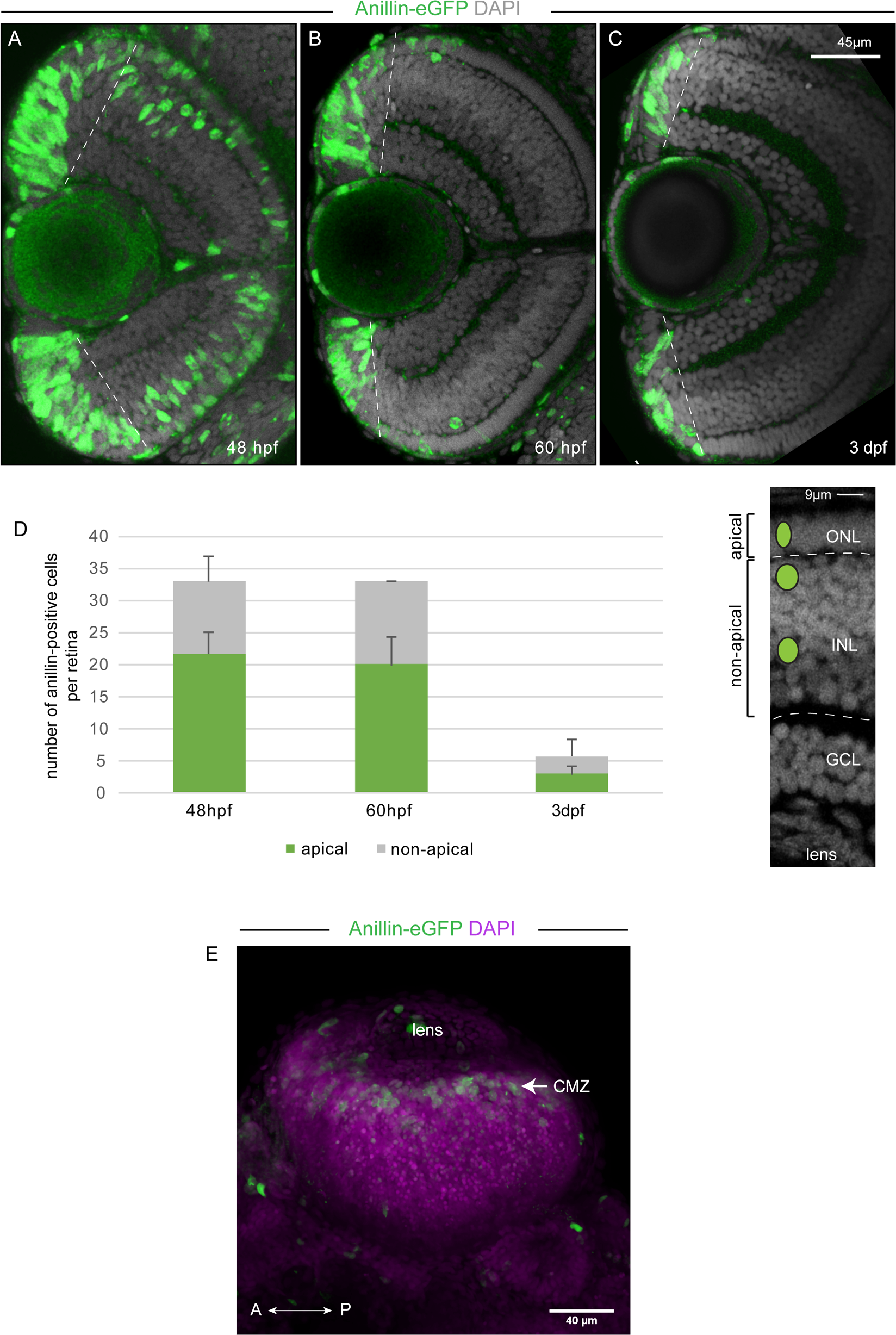
Fig. 5
Anillin-eGFP labelled cell division activity is restricted to the CMZ at 3 dpf and at 5 dpf.
(A-C) Optical section from a z-stack taken in the transversal plane (frontal view as represented in the top panel of Fig 4) through the central retina of an anillin:anillin-eGFP transgenic zebrafish fixed at 48 hpf (A), 60 hpf (B) or 3 dpf (C). Counterstaining with DAPI (grey) marks the cell nuclei and the three retinal cell layers. (D) Left: Anillin-eGFP positive cells were counted, which were allocated to the three retinal nuclear layers of the central retina (excluding the CMZ delineated by the dotted line). Anillin-eGFP positive cells were counted per embryo (48hpf, n = 3 apical = 22±4, non-apical = 11±4; 60 hpf, n = 3, apical = 20±4, non-apical = 13±0; 3dpf, n = 3, apical = 3±3, non-apical = 3±1,5). Right: Overlay of a confocal image with a schematic view representing the different locations of Anillin-eGFP positive cells within the central retina: apical, in the outer nuclear layer (ONL) and non-apical in the inner nuclear layer (INL). Error bars represent standard deviation. GCL: ganglion cell layer. (E) Two-photon z-stack projection (z-sections 1 μm apart) of a retina from a 5 dpf zebrafish showing Anillin-eGFP positive cells (green) in the CMZ. The cell nuclei (magenta) were labelled with DAPI. The image represents a dorsal view.