
|
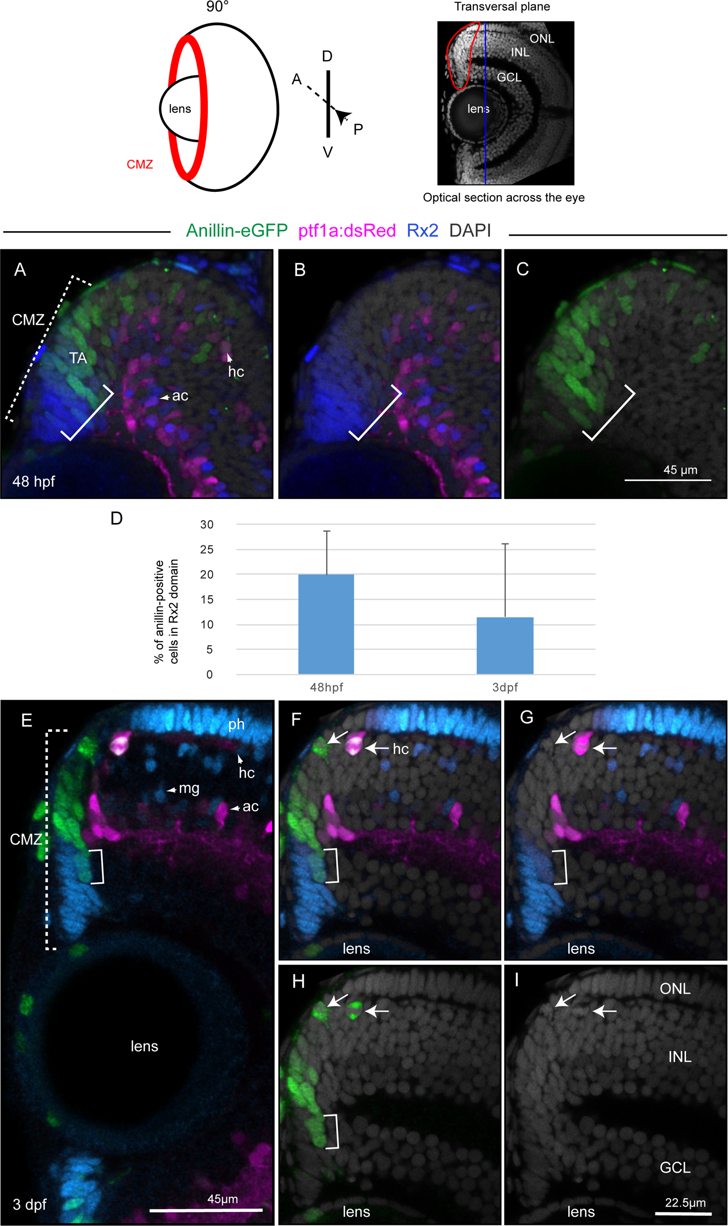
Fig. 4
Anillin-eGFP in the CMZ marks transit-amplifying progenitors entering restricted neuronal lineages.
Top panel: view of the retina in the transversal plane (eye position 90° relative to the viewer). The blue line across the retina corresponds to the sagittal view (optical z-section) in the top panel of Fig 3. (A-C) and (E-I) represent two optical sections from confocal z-stacks through the retina of a 48 hpf (A-C) and 3 dpf (E-I) zebrafish taken in the transversal plane. The squared dotted bracket in (A) and (E) delineates the CMZ domains. The squared bracket in (A-C) and (E-H) delineates cells within the CMZ that are both Anillin-eGFP (green) and Rx2 immunoreactive (blue). (D) As the stem cell niche develops, the percentage of Rx2 immunoreative cells that are also Anillin-eGFP positive in the CMZ domain decreases over time from 20% to 11,4%. The arrows in (F-I) point at two dividing, Anillin-eGFP (green) positive cells and at their nuclei (I). (F,G) One of the two cells is also (ptf1a)dsRed positive and non-apically located. Ph, photoreceptors; ac, amacrine cells; hc, horizontal cells; mg, Müller glial cell. ONL, outer nuclear layer; INL, inner nuclear layer; ONL, outer nuclear layer.