
|
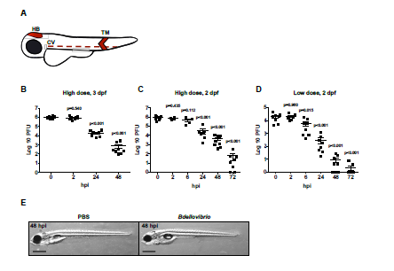
Fig. S1
Injected Predatory Bdellovibrio Persist in Zebrafish Larvae and are Ultimately Cleared, Related to Figure 1
(A) Zebrafish larvae were injected with bacteria or PBS in either the hindbrain ventricle (HB), tail muscle (TM) or caudal vein (CV) shaded in red. Injections in the hindbrain were performed to study population level dynamics of Bdellovibrio, Shigella and/or leukocytes. Injections in the tail muscle were performed to enable highresolution confocal microscopy. (B–D) WT AB zebrafish larvae were injected with (B-C) a high dose of Bdellovibrio or (D) a low dose of Bdellovibrio at (B) 3 dpf or (C-D) 2 dpf in the hindbrain ventricle. Live Bdellovibrio were enumerated from larval homogenates at time points indicated. Each circle represents a count from an individual larva. Mean ± SEM (horizontal bars) are shown. (B) Data pooled from 2 independent experiments using 3-4 larvae per time point. (C) Data from 2 independent experiments using 4 larvae per time point. p values (versus the 0 hpi time point) determined by multiple t-test. Significance with Bonferroni correction defined as p<0.017 for (B), p<0.01 for (C-D). (E) WT AB zebrafish larvae were injected in the hindbrain at 3 dpf with PBS or 1-2 x 10⁵ PFU of Bdellovibrio and imaged via stereomicroscopy at 48 hpi (i.e. 5 dpf). Representative images of larval morphology are shown. Scale bar = 0.5 mm.