
|
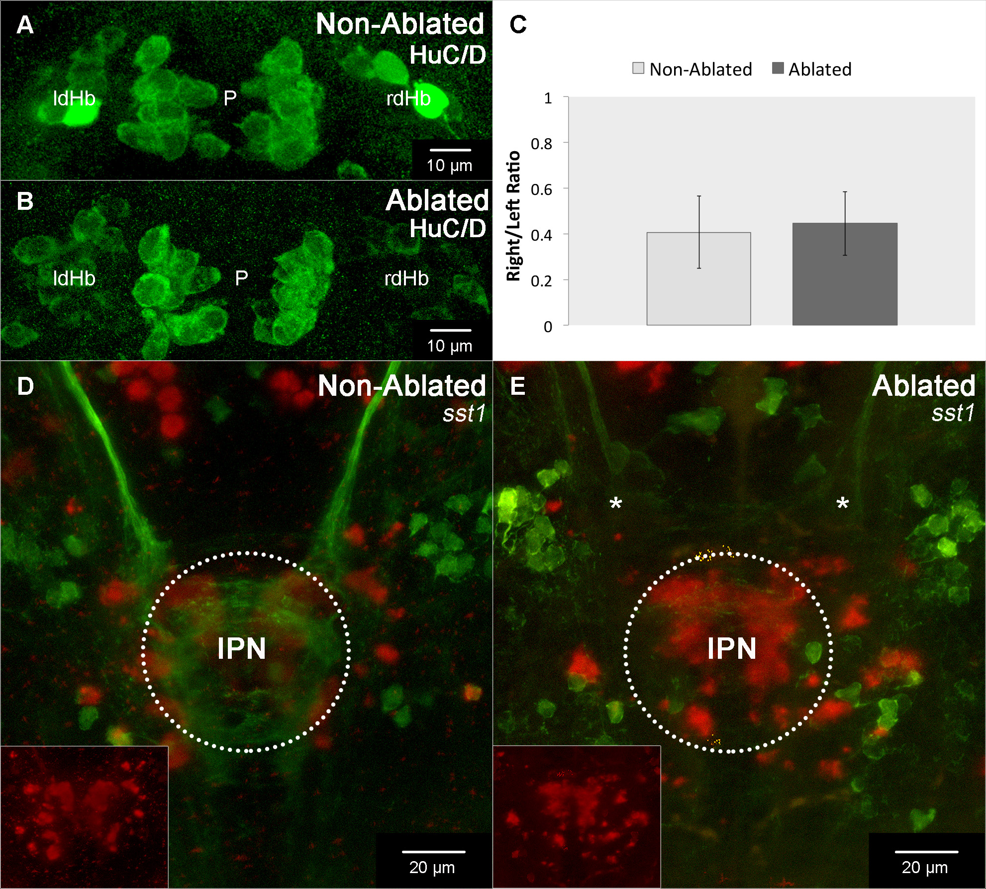
Fig. S4
ThEPC ablations specifically affect dHb network formation.
(A, B, D, E) Dorsal views, anterior to the top, MIPs focussed on (A, B) the habenulae and (D, E) the IPN of 4 dpf embryos. The original stacks were cropped and the gamma adjusted for display purposes. (A-C) The number of HuC/D positive differentiating habenular neurons (non-ablated: n = 9,86 +/- 3,48; ablated: n = 13,14 +/- 5,52; p = 0,32) and their ratio (non-ablated: n = 0,41 +/- 0,16 ; ablated: n = 0,45 +/- 0,14; p = 0,68) between the left and right side is not altered by unilateral ThEPC ablation (n = 7). In addition, also pineal cell development was unaffected (non-ablated: n = 25,12 +/- 3,05; ablated: n = 23,14 +/- 2,54; p = 0,23; n = 7). (D, E) Unilateral ThEPC ablation results in the stalling of dHb efferent axons (asterisks) before reaching the normally formed IPN (encircled) as marked by somatostatin-1 (sst1) expression (red) in Et(-1.0otpa:mmGFP)hd1 embryos. The insets show sst1 expression in the IPN in the red channel. dHb, dorsal habenula; IPN, interpeduncular nucleus; l, left; p, pineal; r, right.