
|
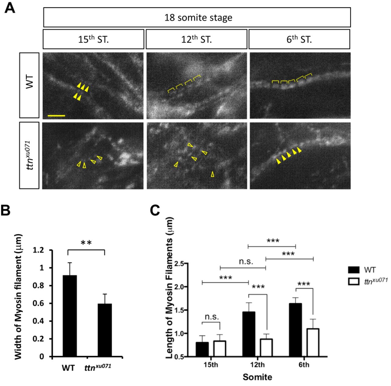
Fig. 7
Phenotypes of thick filament assembly in the ttn.2/1-null mutant. (A) Immunostaining was performed in WT or ttnxu071 mutant embryos at the 18-somite stage with an F59 antibody against the myosin heavy chain. Scale bar: 5 μm. Myosin filaments were aligned longitudinally (solid arrowheads) at the 15th somite in WT embryos but remain scattered in ttnxu071 (open arrowheads). Expansion of thick filaments is seen in WT embryos at the 12th and 6th somites (brackets). However, the thick filaments in the ttnxu071 mutant remain scattered at the 12th somite (open arrowheads) and become aligned at the 6th somite (solid arrowheads). (B) The width of the thick (myosin) filaments of WT and ttnxu071 mutants at the 6th somite was quantified. Means±s.d., N=84 for each group from six embryos. **P<0.01. (C) The length of myosin filaments in the 6th, 12th and 15th somites of WT and ttnxu071 mutant embryos. The length of thick filaments does not increase in the ttnxu071 mutant, whereas thick filament length increases to 1.6 µm in WT. Means±s.d., ***P<0.00001. N=80 for each group in different somites from six embryos. n.s., not significant (P>0.05).