
Image
|
Figure Caption
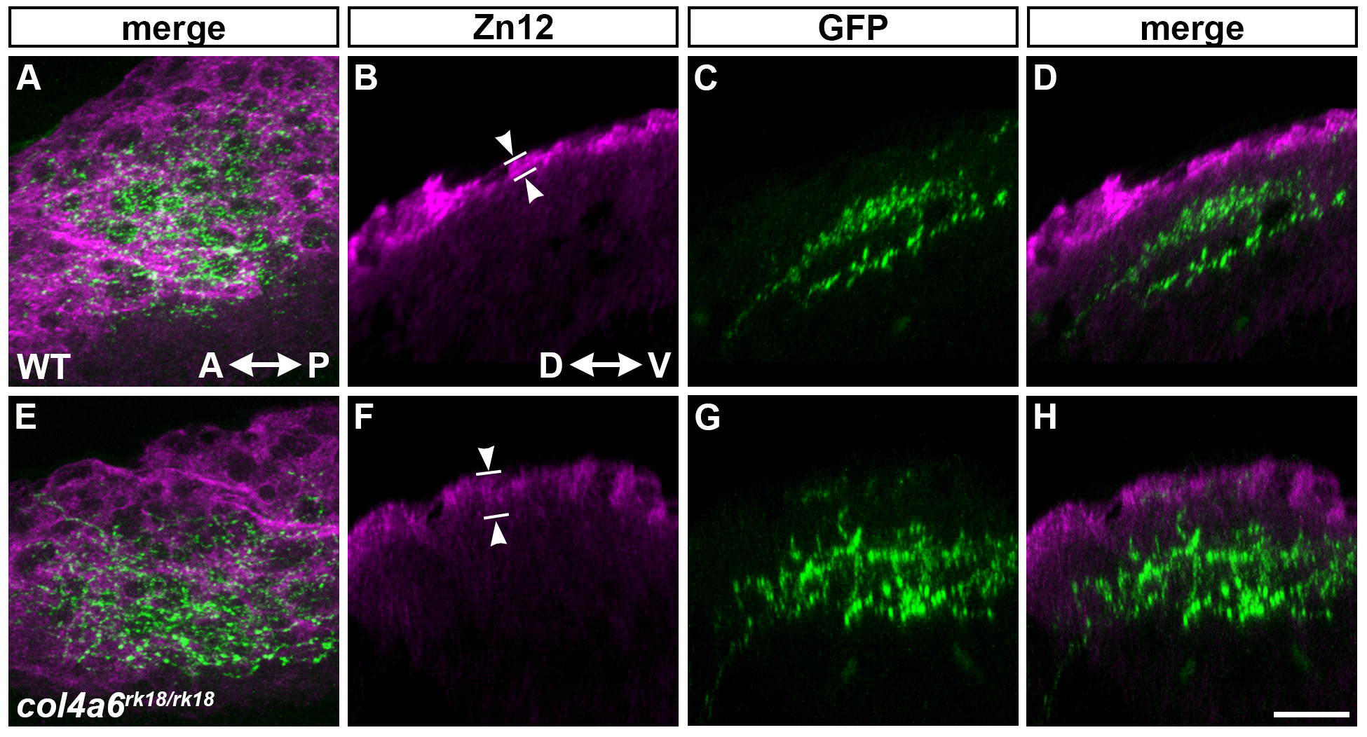
Fig. S6
HNK–1 epitopes are sparsely distributed in the tectal BM region of col4a6 mutants.
Wild-type (A-D) and col4a6 mutant (E-H) larvae were stained with anti-HNK–1 (zn12, A, B, D, E, F, H) and anti-GFP (A, C, D, E, G, H) antibodies. The RGC axons marked by pou4f3:Gal4; UAS:GAP-GFP in the tectal region; dorsal projection views (A, E) and lateral views (B-D, F-H). The HNK–1+ region was thinker in the tectal BM of the col4a6 mutants (n = 5), compared to that in the wild type larvae (n = 3). The statistic analysis is shown inS4 Table. Scale bars: 20 μm in H (applied to A-G).
Acknowledgments
This image is the copyrighted work of the attributed author or publisher, and
ZFIN has permission only to display this image to its users.
Additional permissions should be obtained from the applicable author or publisher of the image.
Full text @ PLoS Genet.