
Image
|
Figure Caption
Fig. S12
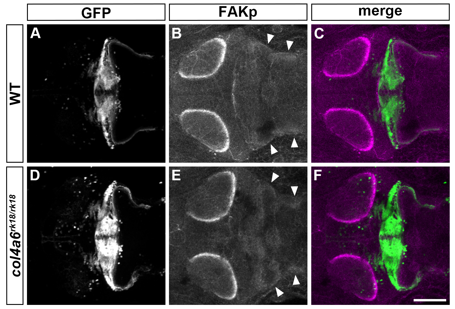
Col4a6-independent FAK activation in GC axons.
Immunostaining of 5-dpf wild-type (A-C) and col4a6 (D-F) mutant larvae harboring the gSA2AzGFF152B; UAS:GFP transgene with anti-GFP (granule cell axons, A, C, D, F) and anti-phosphorylated FAK (B, C, E, F) antibodies. Dorsal views of the rostral hindbrain region. Note that the phosphorylated (active) form of FAK was similarly detected in the caudolateral GC axons in the wild-type and col4a6 mutant hindbrain (marked by arrowheads). Scale bar: 100 μm in F (applied to A-E).
Acknowledgments
This image is the copyrighted work of the attributed author or publisher, and
ZFIN has permission only to display this image to its users.
Additional permissions should be obtained from the applicable author or publisher of the image.
Full text @ PLoS Genet.