
|
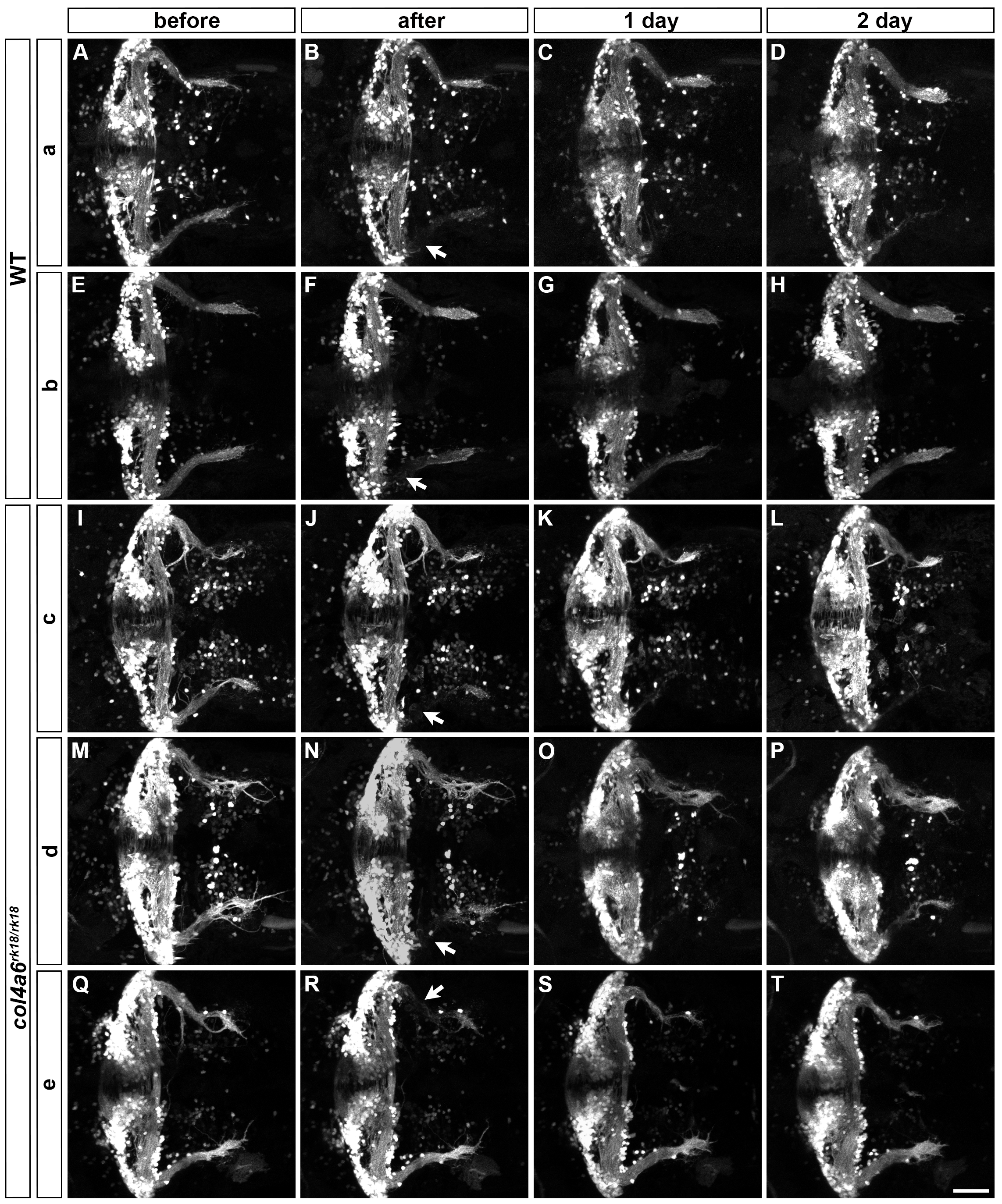
Fig. S9
Laser ablation of GCs.
Axons of caudolateral GCs in wild-type (2 larvae: a, b) and col4a6 mutant (3 larvae: c-e) larvae that harbored hspGFFDMC90A; UAS:Kaede (a, c, d) or hspGFFDMC90A; UAS:GFP (b, e) transgenes were ablated by a laser at 5 dpf. The GC axons of the larvae were observed before (A, E, I, M, Q), soon after (B, F, J, N, R), or 1 (C, G, K, O, S) or 2 days (D, H, L, P, T) after the laser ablation. The experimental conditions were the same as described in the legend for Fig 8. More examples are shown in this figure. Dorsal views of the rostral hindbrain regions. The ablation points are indicated by arrows (B, F, J, N, R). Scale bars: 50 μm in T (applied to A-S).