
|
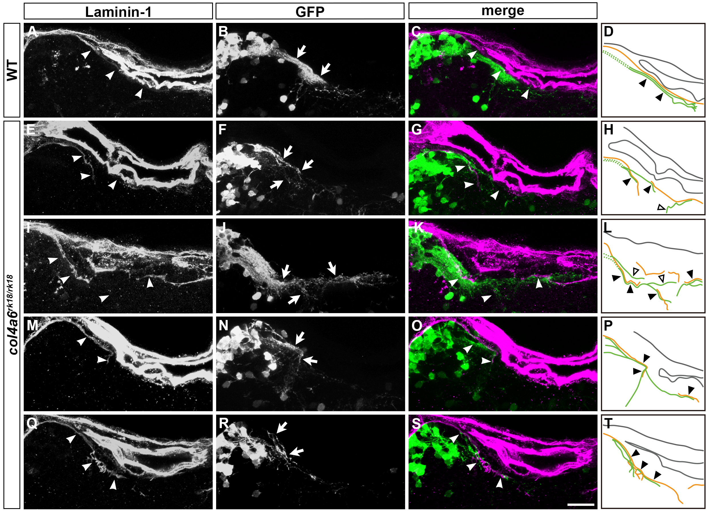
Fig. S8
Abnormal GC axons are coupled with abnormal BM in the col4a6 mutant hindbrain.
The GC axons and BM structures in the hindbrain of wild-type (n = 2) and col4a6 mutant larvae (n = 5) were analyzed as described in Fig 7. Typical examples were shown in Fig 7, and the rest of the samples (one) for WT and four for the mutant) are shown in this figure. Laminin–1 (A, E, I, M, Q), GFP (B, F, J, N, R), and merged images (C, G, K, O, S). Hindbrain BM and caudolateral GC axons are indicated by arrowheads and arrows, respectively. (D, H, L, P, T) Schematic representation of the BM (brown) and GC axons (green). GC axons that ran along the BM are indicated by closed triangles. GC axons that did not run along the BM are indicated by open triangles. Scale bars: 20 μm in S (applied to A-C, E-G, I-K, M-O, Q-R).