
Image
|
Figure Caption
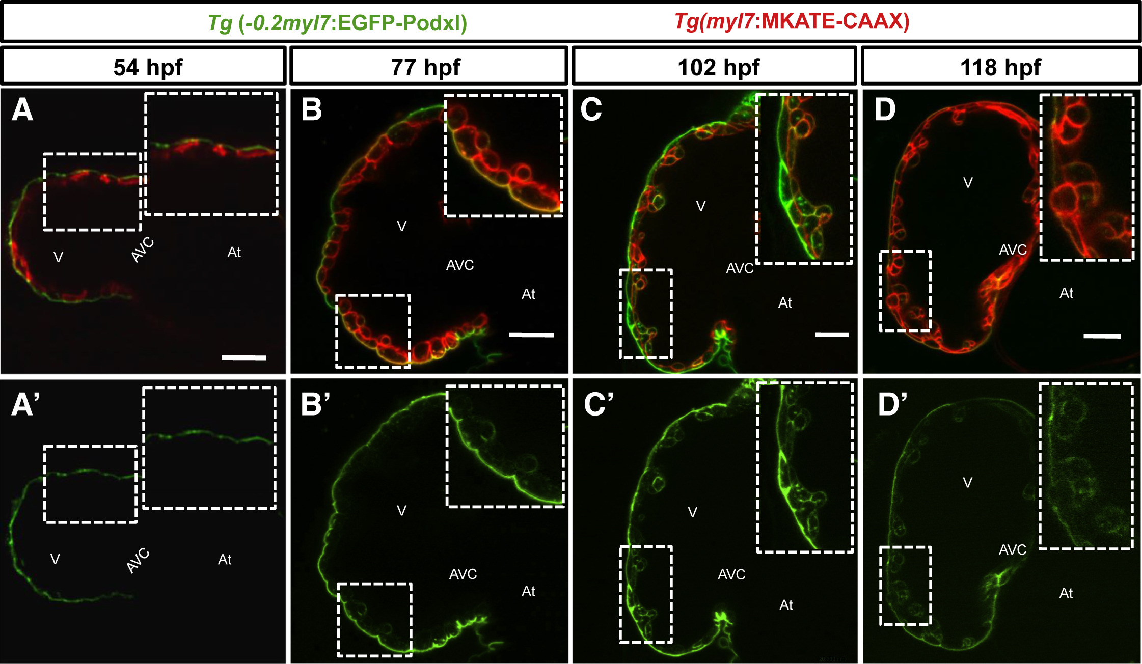
Fig. 2
EGFP-Podocalyxin Localization in CMs during Cardiac Trabeculation
(A–D') Confocal images (mid-sagittal sections) of Tg(−0.2myl7:EGFP-podxl);Tg(myl7:MKATE-CAAX) zebrafish hearts at 54 (A and A'), 77 (B and B'), 102 (C and C'), and 118 (D and D′) hpf. Boxed areas show high-magnification images of MKATE-CAAX and/or EGFP-Podxl expression (A–D'). Compact-layer CMs remain polarized at least until 118 hpf. At, atrium; V, ventricle; AVC, atrioventricular canal. Scale bars, 20 μm.
Figure Data
Acknowledgments
This image is the copyrighted work of the attributed author or publisher, and
ZFIN has permission only to display this image to its users.
Additional permissions should be obtained from the applicable author or publisher of the image.
Full text @ Cell Rep.