
|
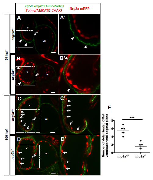
Fig. S1
Lower number of delaminated cardiomyocytes in nrg2a mutants
(A-D') Confocal images (mid-sagittal sections) of Tg(-0.2myl7:EGFP-podxl);Tg(myl7:MKATECAAX) zebrafish hearts from a nrg2a+/- incross at 52 (A-B') and 100 (C-D') hpf . At 52 hpf, only polarized cardiomyocytes are found in both nrg2a+/- (A and A') and nrg2a-/- (B and B') embryos. Arrowheads point to apical localization of EGFP-Podxl. At 100 hpf, while a high number of delaminated cardiomyocytes are observed in nrg2a+/- larvae (C and C', white arrows), there are only a few delaminated cardiomyocytes in nrg2a-/- larvae (D and D' arrows). (E) Graph showing the number of delaminated cardiomyocytes in 100 hpf nrg2a heterozygous and homozygous mutant larvae, n=5 each. Each dot represents one heart. Data are shown as mean ± SEM. *** P< 0.001 by Student's t-test. At, atrium; V, ventricle; AVC, atrioventricular canal. Scale bars, 20 μm.