
Fig. S1 H
- ID
- ZDB-IMAGE-170103-24
- Genes
- Publication
- Qi et al., 2016 - NgAgo-based fabp11a gene knockdown causes eye developmental defects in zebrafish
- All Figures
- Figures for Qi et al., 2016
|
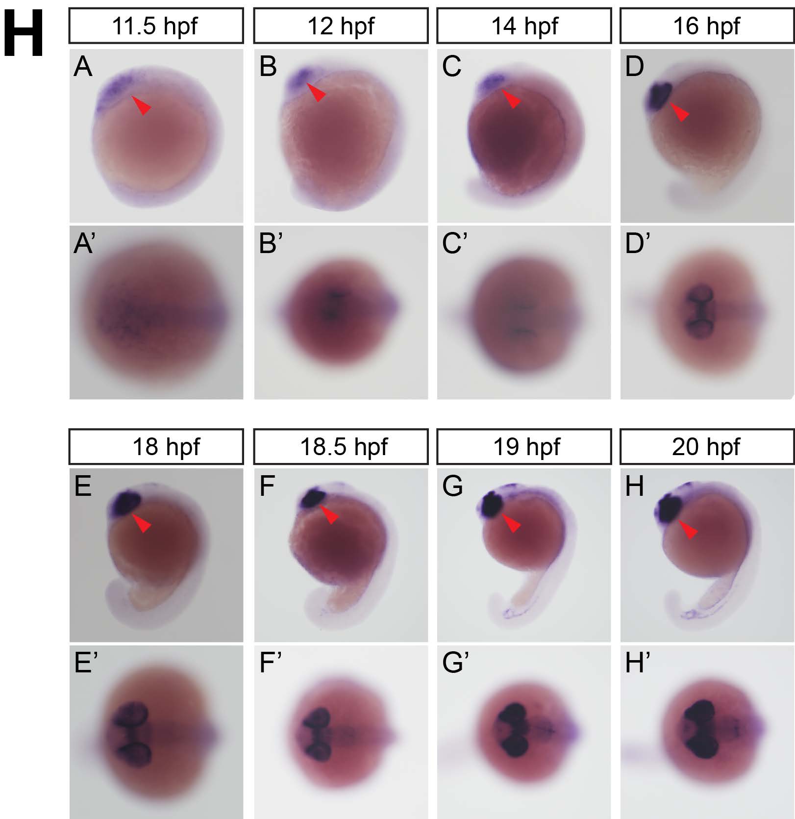
Fig. S1 H
Whole mount in situ hybridization analysis of zebrafish embryos using antisense fabp11a probe. (A) 11.5 hpf, lateral view. Red arrowhead indicates eye field. (A'). 11.5 hpf, coronal view. (B) 12 hpf, lateral view. Red arrowhead indicates eye field. (B'). 12 hpf, coronal view. (C) 14 hpf, lateral view. Red arrowhead indicates eye field. (C'). 14 hpf, coronal view. (D) 16 hpf, lateral view. Red arrowhead indicates eye field. (D'). 16 hpf, coronal view. (E) 18 hpf, lateral view. Red arrowhead indicates eye field. (E'). 18 hpf, coronal view. (F) 18.5 hpf, lateral view. Red arrowhead indicates eye field. (F'). 18.5 hpf, coronal view. (G) 19 hpf, lateral view. Red arrowhead indicates eye field. (G'). 19 hpf, coronal view. (H) 20 hpf, lateral view. Red arrowhead indicates eye field. (H'). 20 hpf, coronal view.