
|
Fig. S1
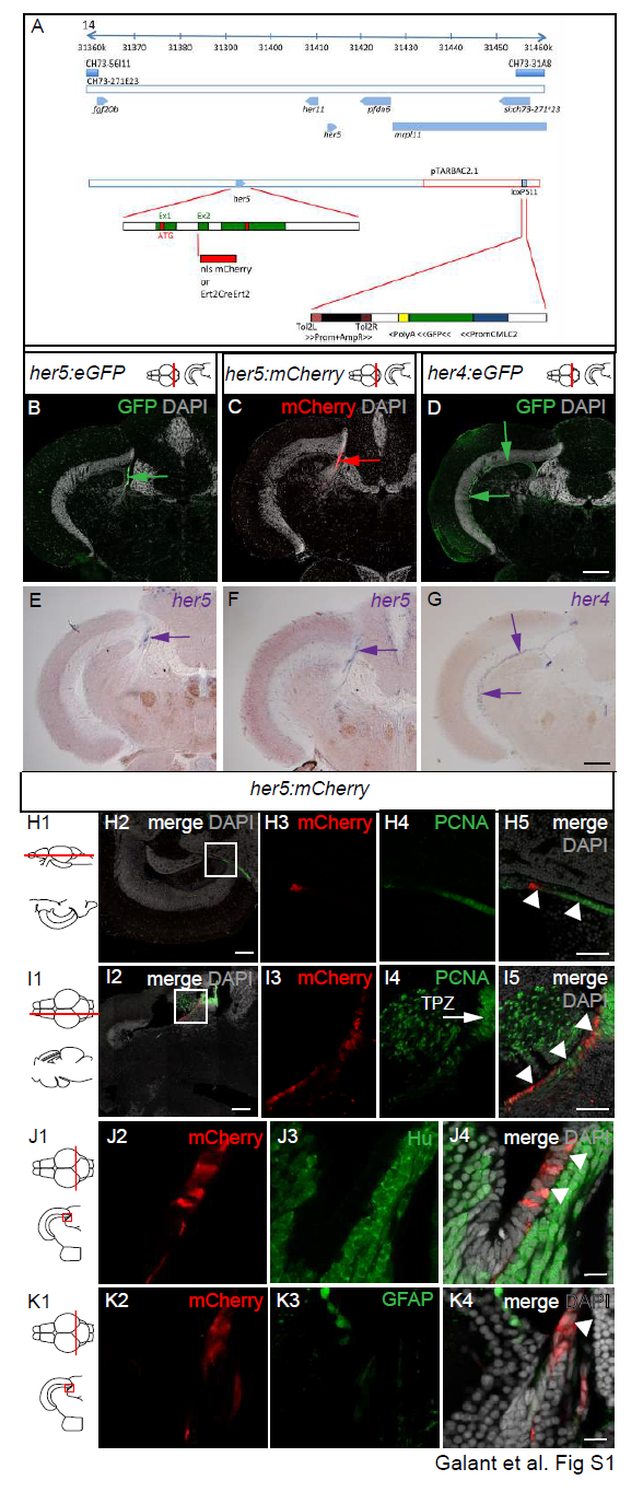
Construction and expression profile of Tg(her5:mCherry) compared with the expression of other transgenic reporters, genes and cell type markers in the adult midbrain. A. Schematic of the genomic area covered by the her5-containing BAC (pTARBAC2.1) used for transgenesis. The her5 locus is magnified in green (exons) and white (untranslated areas), and red domains depicts the start and stop codons. The mCherry or Ert2CreErt2 cassettes (red box) were inserted in frame within the second exon of her5. The entire BAC was flanked with Tol2 sites and used for transgenic generation, these sequences coupled with a cmlc2:GFP cardiac cassette (blue and green box) were inserted at the loxP511 locus. B-D. Compared reporters expression in Tg(her5:eGFP) (Tallafuss and Bally-Cuif, 2003), Tg(her5:mCherry) (this study) and Tg(her4:eGFP) (Yeo et al., 2007) transgenic lines in the adult midbrain. Cross sections at the level indicated on the schematics, counterstained with DAPI; arrows point to expression sites. E-G. In situ hybridization for the endogenous genes (blue signal, blue arrows) in the different transgenic lines. H,I. Compared expression of Her5- mCherry with PCNA on horizontal (H) and sagittal (I) sections (planes indicated on the schematics) of an adult midbrain observed in confocal microscopy. Panels 2: low magnification views, with merged fluorescence channels and counterstained with DAPI. Panels 3-5: high magnifications of the area boxed in panels 2, channels/stainings colorcoded. J, K. Compared expression of Her5-mCherry with the neuronal marker HuC/D (J) and the glial marker GFAP (K), respectively, on cross sections of an adult midbrain observed in confocal microscopy. Panel 3-4: High magnifications of the area boxed in the schematics, with channels/stainings color-coded. White arrowheads to the PML on panels H,I5 and J,K4. Scale bars: B-G 50μm, H,I 100μm, J-K 10μm. Abbreviations: TPZ: tectal proliferation zone.
Reprinted from Developmental Biology, 420(1), Galant, S., Furlan, G., Coolen, M., Dirian, L., Foucher, I., Bally-Cuif, L., Embryonic origin and lineage hierarchies of the neural progenitor subtypes building the zebrafish adult midbrain, 120-135, Copyright (2016) with permission from Elsevier. Full text @ Dev. Biol.