
|
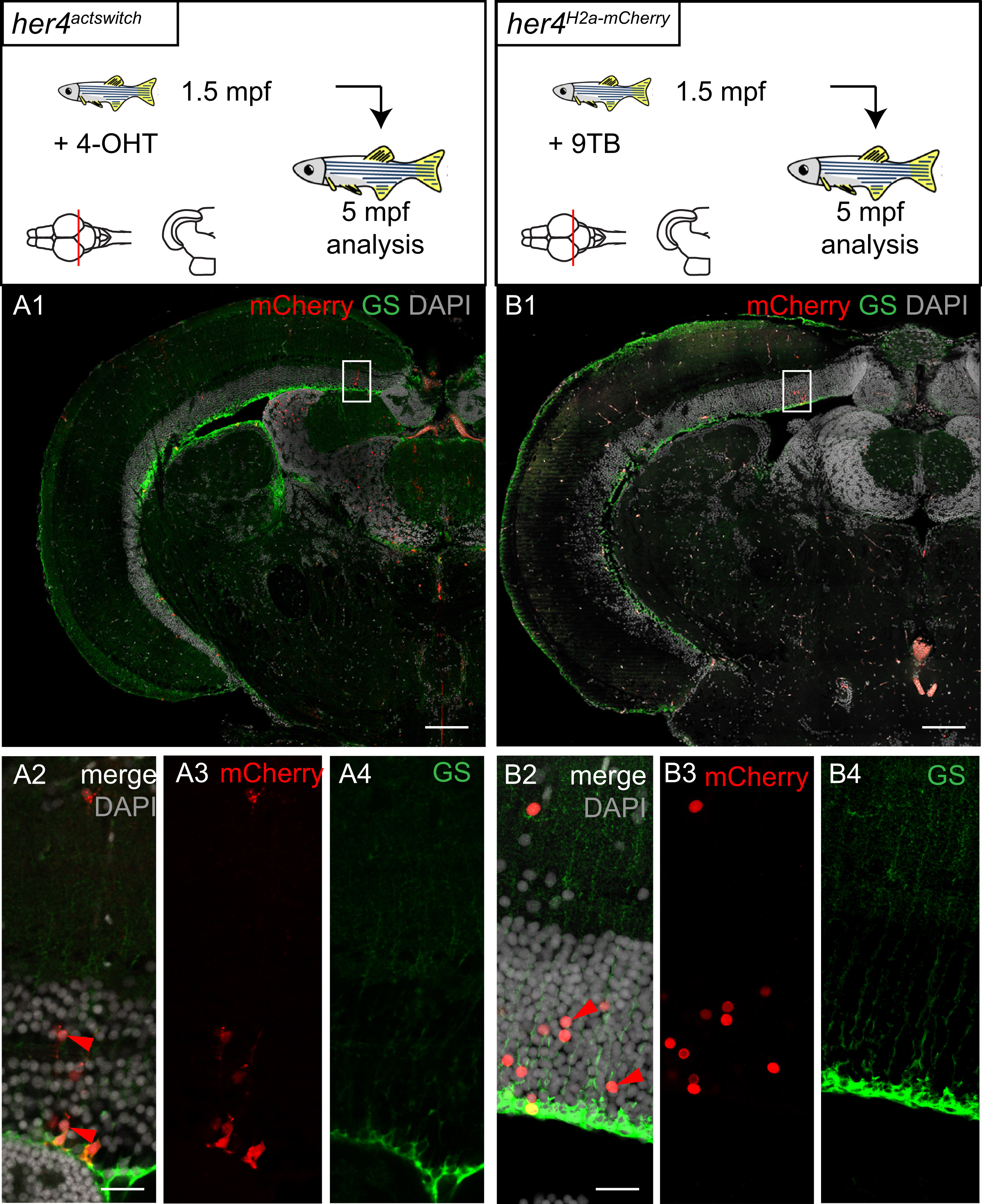
Fig. 4
her4-positive RG exhibit neurogenic activity in the post-embryonic TeO. Compared fate in the 5mpf TeO of RG cells expressing her4 at 1.5mpf, assessed by Cre-mediated tracing (A) and rtTA-mediated birthdating (B). Panels 1. Cross-sections of a TeO hemisphere immunostained for mCherry and GS and counterstained with DAPI and observed in confocal microscopy. Panels 2–4. High magnifications of the areas boxed in panels 1 (single and merged channels as indicated, maximum projection of 4 sections over 3 µm). In both cases, a single stripe of mCherry-positive progeny cells is observed, identically located along the antero-posterior and dorso-ventral axes and comprising both neurons and RG. Scale bars: A1 and B1 100 µm, A2-A4 and B2-B4 50 µm.
Reprinted from Developmental Biology, 420(1), Galant, S., Furlan, G., Coolen, M., Dirian, L., Foucher, I., Bally-Cuif, L., Embryonic origin and lineage hierarchies of the neural progenitor subtypes building the zebrafish adult midbrain, 120-135, Copyright (2016) with permission from Elsevier. Full text @ Dev. Biol.