
|
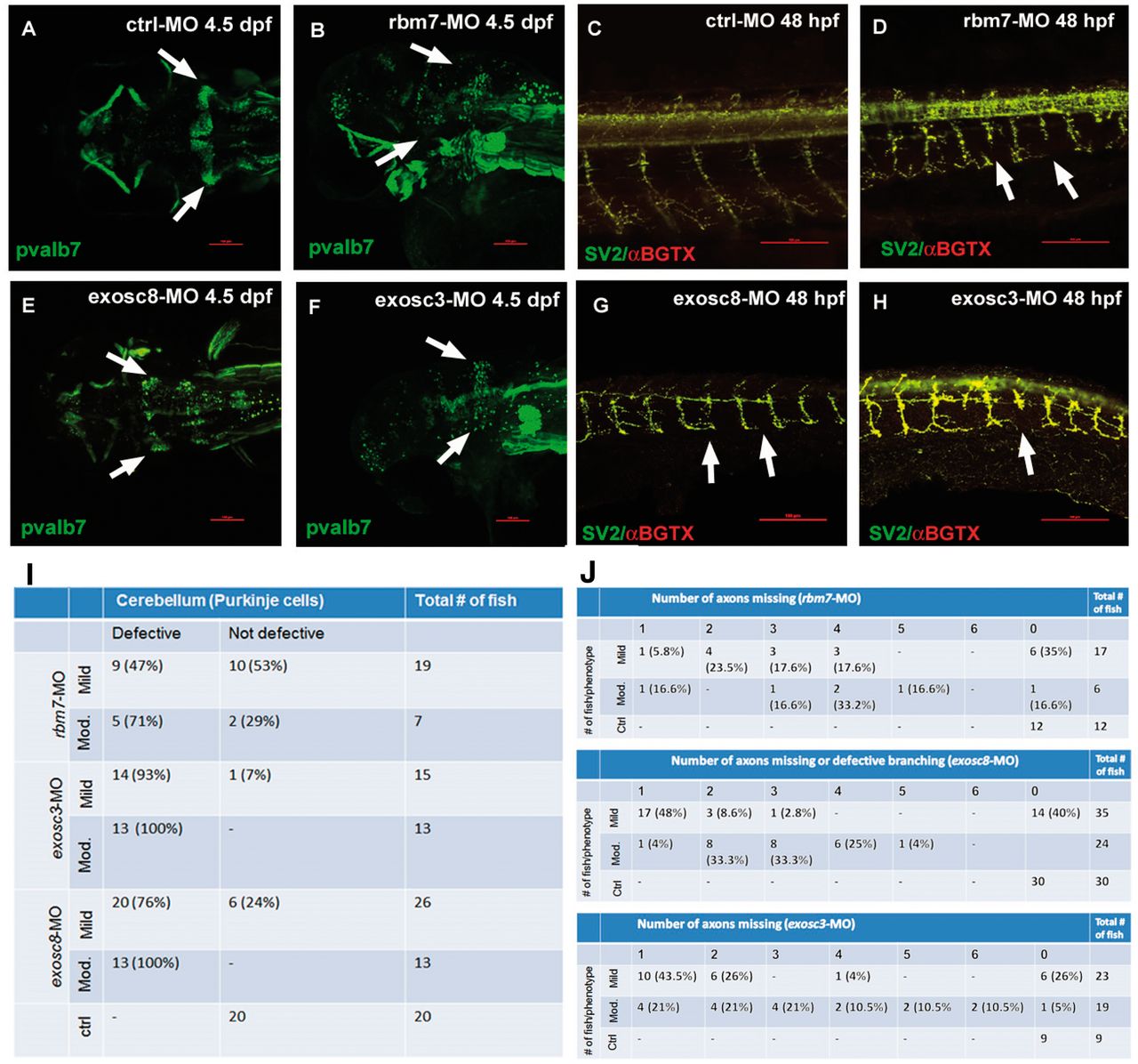
Fig. 5
Confocal microscopy analysis of PCs (A,B,E,F) and neuromuscular junctions (C,D,G,H) in zebrafish upon injection of three different MOs. PCs fail to differentiate in rbm7-MO (B), exosc8-MO (E) and exosc3-MO (F) zebrafish compared with ctrl-MO fish (A). The percentage of fish with cerebellar defects varies significantly between morphants, reflecting what observed in patients. Motor axon growth is defective in all three morphants (D,G,H); arrows point at abnormally short motor axons. Interestingly, only in exosc8-MO the motor neuron axon branches in close proximity of the spinal cord instead of more ventrally as in the ctrl-MO fish (G). Scale bar = 100 μm. We show the quantity and respective percentage of fish with cerebellar defects (I). Axonal defects in different morphant and phenotypical classes (only mild and moderate phenotypes were considered for this analysis) (J). The number of defective structures increases with the severity of the phenotype.